. 2019 Sep:26:101274.
doi: 10.1016/j.redox.2019.101274. Epub 2019 Jul 10.

8-Chloroadenosine induces apoptosis in human coronary artery endothelial cells through the activation of the unfolded protein response

Affiliations

8-Chloroadenosine induces apoptosis in human coronary artery endothelial cells through the activation of the unfolded protein response

Vickie Tang et al. Redox Biol. 2019 Sep.

Abstract

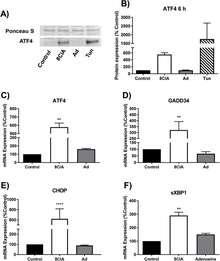
Infiltration of leukocytes within the vessel at sites of inflammation and the subsequent generation of myeloperoxidase-derived oxidants, including hypochlorous acid, are key characteristics of atherosclerosis. Hypochlorous acid is a potent oxidant that reacts readily with most biological molecules, including DNA and RNA. This results in nucleic acid modification and the formation of different chlorinated products. These products have been used as biomarkers of inflammation, owing to their presence in elevated amounts in different inflammatory fluids and diseased tissue, including atherosclerotic lesions. However, it is not clear whether these materials are simply biomarkers, or could also play a role in the development of chronic inflammatory pathologies. In this study, we examined the reactivity of different chlorinated nucleosides with human coronary artery endothelial cells (HCAEC). Evidence was obtained for the incorporation of each chlorinated nucleoside into the cellular RNA or DNA. However, only 8-chloro-adenosine (8ClA) had a significant effect on the cell viability and metabolic activity. Exposure of HCAEC to 8ClA decreased glycolysis, and resulted in a reduction in ATP, with a corresponding increase in the chlorinated analogue, 8Cl-ATP in the nucleotide pool. 8ClA also induced sustained endoplasmic reticulum stress within the HCAEC, which resulted in activation of the unfolded protein response, the altered expression of antioxidant genes and culminated in the release of calcium into the cytosol and cell death by apoptosis. Taken together, these data provide new insight into pathways by which myeloperoxidase activity and resultant hypochlorous acid generation could promote endothelial cell damage during chronic inflammation, which could be relevant to the progression of atherosclerosis.

Keywords: DNA; Hypochlorous acid; Inflammation; Myeloperoxidase; Nucleoside; RNA.


Figures

Image 1
Graphical abstract
Fig. 1
Fig. 1
The incorporation of chlorinated nucleosides into cellular RNA and DNA. HCAEC (1 × 106 cells) were treated with A) 8ClA, B) 8ClG, C) 5ClC, D) 8CldA, E) 8CldG and F) 5CldC (16 μM; 0.16 pmol/cell) for 2–72 h (grey bars) or 24 h with washing to remove exogenous chlorinated nucleosides and re-incubation of the cells in normal cell media for 48 h (white bars, labelled 24 + 48), before extraction, hydrolysis and dephophorylation of the RNA and DNA and analysis by LC-MS. The concentration of chlorinated nucleoside was normalised to the respective parent nucleoside and is expressed as a 1:1000 ratio. Data are the mean ± SEM samples from at least three independent cell donors. *, ** and **** represent a significant (p < 0.05, 0.01 and 0.0001) difference in the level of chlorinated nucleosides compared to the respective non-treated controls, with a, b, and c showing a significant (p < 0.05) difference on comparing the 24 + 48 sample with the 24 h, 48 h or 72 h samples respectively, as determined by 1-way ANOVA with the Sidak multiple comparison post-hoc test.
Fig. 2
Fig. 2
The effect of chlorinated nucleosides on metabolic activity. HCAEC (1 × 104 cells) were treated each chlorinated nucleoside (4, 8, 16 μM) for A) 24 h or B) 48 h before assessing metabolic activity by conversion of the resazurin-based PrestoBlue® reagent to fluorescent resorufin at λex = 560 nm and λem = 590 nm. Results represent the mean ± SEM of multiple experiments with at least 3 independent cell donors. * and ** show a significant (p < 0.05 and 0.01) change in metabolic activity compared to the non-treated control by two-way ANOVA with the Dunnett's multiple comparison post-hoc test.
Fig. 3
Fig. 3
Treatment of HCAEC with 8ClA results in 8Cl-ATP formation. (A) and (B) HCAEC (6 × 105 cells) were treated with 8ClA (8 μM) or adenosine (8 μM) for 24 h before quantifying the concentration of ATP (A) and 8Cl-ATP (B) in the cellular nucleotide pool following extraction with perchloric acid and separation by HPLC. (C) HCAEC (1 × 104 cells) were treated with 8ClA (4–16 μM) or adenosine (16 μM) for 48 h with ATP quantified using the ATPlite assay. Data represent the mean ± SEM of multiple replicates with at least 2 independent cell donors. * shows a significant (p < 0.05) change compared to the non-treated control samples by one-way ANOVA with Dunnett's multiple comparison post-hoc test.
Fig. 4
Fig. 4
The effect of 8ClA on mitochondrial respiration. The change in mitochondrial respiration of HCAEC (3.2 × 104 cells) treated with 8ClA (4, 8, 16 μM) or adenosine (16 μM) for 48 h was determined by measuring the OCR using a Seahorse XF Analyser with the sequential addition of oligomycin (1.7 μM), FCCP (0.7 μM) and rotenone/antimycin A (0.5 μM). The results represent the mean ± SEM of 4 biological replicates with 3 independent cell donors.
Fig. 5
Fig. 5
The effect of 8ClA on glycolysis and GAPDH activity. HCAEC (3.2 × 104 cells) were treated with 8ClA (4, 8, 16 μM) or adenosine (16 μM) for 48 h before assessing: A,B) basal glycolytic rate using a Seahorse XF analyser to measure ECAR following injection of glucose (10 mM), C) the concentration of extracellular l-lactate and D) GAPDH activity. The results represent the mean ± SEM of 4 biological replicates with 3 independent cell donors. * (p < 0.05), ** (p < 0.01), *** (p < 0.001), **** (p < 0.0001) represent a significant decrease compared to control samples analysed using a one-way ANOVA with Dunnett's multiple comparison post-hoc test.
Fig. 6
Fig. 6
8ClA treatment induces ER stress and activation of the UPR. HCAEC (1 × 105 cells) were treated with 8ClA or adenosine (8 μM) for 6 h (panel A) or 24 h (panels B–E) before assessing markers of ER stress. (A,B) Western blot analysis of nuclear extracts showing translocation of ATF4 from the cytosol into the nucleus compared to treatment with tunicamycin (Tun; 2.5 μg/mL). Graphs (C–F) show the mRNA expression of ATF4, GADD34, CHOP and sXPB1 at 24 h, as analysed by qRT-PCR with normalisation to the expression of the housekeeping genes 18S and RPL13. Results represent the mean ± SEM of 3 biological replicates with 3 independent cell donors and are expressed as an increase relative to the non-treated control. ** (p < 0.01) and **** (p < 0.0001) represent a significant increase compared to control using a one-way ANOVA with Dunnett's multiple post-hoc comparisons test.
Fig. 7
Fig. 7
8ClA alters the expression of cellular antioxidant responses in HCAEC. HCAEC (1 × 105 cells) were treated with 8ClA or adenosine (8 μM) for 24 h before assessing the expression of antioxidant genes or proteins. Graphs (A–D) show the mRNA expression of NQO1, HO-1, GCLC and TXNIP at 24 h, as analysed by qRT-PCR with normalisation to the expression of the housekeeping genes 18S and RPL13. Panels E and F show the alteration in expression of TXNIP protein by Western blotting, normalised to the expression of β-actin. Results represent the mean ± SEM of 3 biological replicates with 3 independent cell donors and are expressed as an increase relative to the non-treated control. * (p < 0.05) and ** (p < 0.01) represent a significant increase compared to control using a one-way ANOVA with Dunnett's multiple post-hoc comparisons test.
Fig. 8
Fig. 8
8ClA changes the distribution of intracellular calcium and promotes apoptotic cell death in HCAEC. HCAEC (1 × 105 cells) were treated with 8ClA or adenosine (8 μM) before assessing A,B) changes in cytosolic calcium with and without pre-treatment of the HCAEC with the PERK inhibitor GSK2606414 (10 μM) following 24 h treatment with 8ClA using Fluo-4AM. C) Cell viability following 48 h and D) 72 h treatment with 8ClA using Annexin V and propidium iodide staining, with flow cytometry analysis in each case. Results represent the mean ± SEM of 3 biological replicates with 3 independent cell donors. * (p < 0.05), ** (p < 0.01), *** (p < 0.001) and **** (p < 0.0001) represent a significant increase compared to control using a two-way ANOVA with Dunnett's multiple post-hoc comparisons test.

Similar articles

Cited by

References

    1. Libby P. Inflammation in atherosclerosis, arterioscler. Thromb. Vasc. Biol. 2012;32(9):2045–2051. - PMC - PubMed
    1. Davies M.J., Hawkins C.L., Pattison D.I., Rees M.D. Mammalian heme peroxidases: from molecular mechanisms to health implications. Antioxidants Redox Signal. 2008;10(7):1199–1234. - PubMed
    1. Rayner B.S., Love D.T., Hawkins C.L. Comparative reactivity of myeloperoxidase-derived oxidants with mammalian cells. Free Radic. Biol. Med. 2014;71:240–255. - PubMed
    1. Teng N., Maghzal G.J., Talib J., Rashid I., Lau A.K., Stocker R. The roles of myeloperoxidase in coronary artery disease and its potential implication in plaque rupture. Redox Rep. 2017;22(2):51–73. - PMC - PubMed
    1. Zhang R., Brennan M.L., Fu X., Aviles R.J., Pearce G.L., Penn M.S., Topol E.J., Sprecher D.L., Hazen S.L. Association between myeloperoxidase levels and risk of coronary artery disease. J. Am. Med. Assoc. 2001;286(17):2136–2142. - PubMed

Publication types

MeSH terms