. 2019 Aug;8(8):1136-1148.
doi: 10.1530/EC-19-0242.

N-Glycosylation influences human corticosteroid-binding globulin measurements

Affiliations

N-Glycosylation influences human corticosteroid-binding globulin measurements

Lesley A Hill et al. Endocr Connect. 2019 Aug.

Abstract

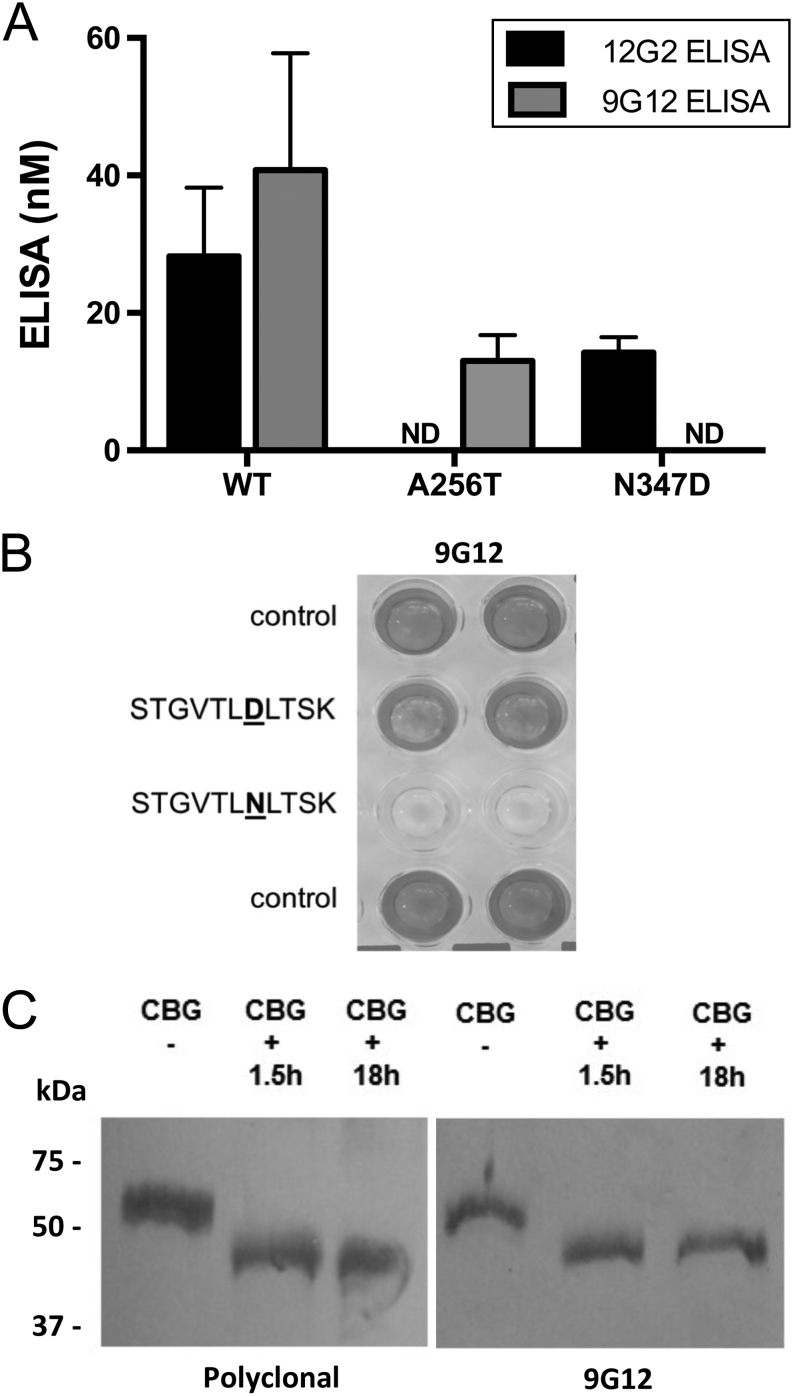
Objective: Discrepancies in ELISA measurements of human corticosteroid-binding globulin (CBG) using detection monoclonal antibodies that recognize an epitope (9G12) within its reactive center loop (RCL), versus an epitope (12G2) in a different location, have suggested that CBG with a proteolytically cleaved RCL exists in blood samples. We have previously been unable to verify this biochemically, and sought to determine if N-glycosylation differences account for discrepancies in ELISA measurements of CBG.

Methods and subjects: Molecular biological, biochemical and glycopeptide analyses were used to examine how N-glycosylation at specific sites, including at N347 within the RCL, affect CBG ELISA or steroid-binding capacity assay (BCA) measurements. Plasma from patients with congenital disorders of glycosylation (CDG) was also examined in these assays as examples of N-glycosylation defects.

Results: We demonstrate that an N-glycan at N347 within the CBG RCL limits the 9G12 antibody from recognizing its epitope, whereas the 12G2 antibody reactivity is unaffected, thereby contributing to discrepancies in ELISA measurements using these two antibodies. Qualitative differences in N-glycosylation at N238 also negatively affect the steroid-binding of CBG in the absence of an N-glycan at N347 caused by a T349A substitution. Desialylation increased both ELISA measurements relative to BCA values. Similarly, plasma CBG levels in both ELISAs were much higher than BCA values in several CDG patients.

Conclusions: Plasma CBG measurements are influenced by variations in N-glycosylation. This is important given the increasing number of CDG defects identified recently and because N-glycosylation abnormalities are common in patients with metabolic and liver diseases.

Keywords: congenital disorders of glycosylation; epitopes; glycosylation; monoclonal antibodies; protein structure; steroid binding.


Figures

Figure 1
Figure 1
Positions of N-glycans attached to human CBG and the CBG N-glycosylation mutants investigated, the CBG reactive center loop (RCL) sequence and 9G12 monoclonal antibody epitope. (A) Structural model demonstrating the extent of N-linked glycosylation of human CBG in its relaxed (R) conformation. In the R conformation, the CBG RCL has been cleaved and inserted into the core of the protein. The N-glycans shown are based on their reported most frequent compositions (11). The predicted N-glycan at Asn9 could not be added because this position is lacking in the crystal structure that the model is based on. The CBG protein is colored light gray and glycans are colored dark gray, with the exception of those attached to N238 and N347, which are colored black. The steroid-binding site (dashed circle) is indicated, as are epitopes (black) recognized by monoclonal antibodies used in the ELISAs, i.e. A256 for the 12G2 ELISA and S341-L348 for the 9G12 ELISA, in the lower panel. In this representation, RCL-cleaved human CBG (PDB ID 4BB2) was utilized for in silico modeling of N-linked glycans, using the program GlyProt (http://glycosciences.de) and illustrations were created using the program PyMol (http://pymol.org). (B) Positions of the N-glycosylation sites in WT human CBG and in the CBG N-glycosylation mutants studied, including CBG T349A that disrupts glycosylation at N347 within the RCL without directly affecting the 9G12 epitope. (C) The CBG RCL sequence with the N-glycosylation (NLT) sequon in bold. The 9G12 antibody epitope is underlined with an arrow.
Figure 2
Figure 2
N347 is a key recognition amino acid in the epitope for the RCL monoclonal antibody 9G12. (A) WT human CBG is recognized in both the 12G2 and 9G12 ELISAs, whereas the N347D mutant is undetectable using 9G12. The CBG A256T mutant served as a control as it is recognized by the 9G12 antibody and not the 12G2 antibody. Data are expressed as mean ± s.d. for n = 3 technical replicates. (B) Chromogenic signals in horizontal duplicates in microtitre plate wells with RCL antibody 9G12 show immobilized RCL synthetic peptide is recognized and not displaced by either the control (buffer only) or D347 synthetic peptide (STGVTLDLTSK) at 100 µg/mL. This contrasts with complete displacement using N347 RCL peptide (STGVTLNLTSK) at 100 µg/mL. (C) PNGase F treatment (+) of native CBG for 1.5 h or 18 h followed by SDS-PAGE and Western blotting with 9G12 or polyclonal antibodies show equivalent signals and mobility shifts compared to no treatment (−). This indicates some carbohydrate removal but not at N347 as deamidation to D347 would be an expected consequence with the absence of recognition by antibody 9G12. Positions of molecular weight markers (kDa) are on the left.
Figure 3
Figure 3
Loss of N347 glycosylation within the CBG RCL increases 9G12 ELISA values, irrespective of the types of N-glycosylation. (A) Examples of the type of N-glycosylation that can be expected to be attached to CBG expressed in fully glycosylation-competent (CHO-S) and partially deficient (Lec1 and Lec2) CHO cells (45). Lec1 cells lack a functional N-acetylglucosaminyltransferase-I, leading to homogenous high mannose N-glycosylation, whereas Lec2 cells lack the CMP-sialic acid Golgi transporter and are therefore unable to sialylate the expressed glycoproteins. (B) Differences in N-glycosylation on CBG influence its mobility during SDS-PAGE as assessed by Western blotting. The CBG steroid-binding capacity (nM) as measured by BCA is shown for WT CBG and the CBG glycosylation mutants applied to the Western blot. Molecular weight markers in kDa are on the left of the blot. (C) The WT CBG and glycosylation-deficient CBG mutants produced by CHO-S, Lec1 and Lec2 cells were measured using the 12G2 and 9G12 ELISA methods. The CBG concentrations in culture media were higher with the 9G12 ELISA when compared to the 12G2 ELISA. Abnormally high 9G12/12G2 ELISA ratios were observed for the CBG T349A mutant. Data are expressed as mean ± s.d. for n = 3 technical replicates.
Figure 4
Figure 4
Desialylation increases the recognition of plasma CBG in the 12G2 and 9G12 ELISAs. (A) Comparison between the steroid-binding capacity assay (BCA) and 12G2 or 9G12 ELISA measurements of CBG in representative plasma samples in which the two ELISA measurements were either concordant or discrepant. (B) Western blotting of the same plasma samples as in (A) shows that desialylation of CBG N-glycans increased the electrophoretic mobility of CBG. Plasma samples were either untreated (−) or treated (+) with sialidase A. Molecular weight markers in kDa are on the left of the blot. (C) Plasma CBG concentrations were measured by BCA, 12G2 and 9G12 ELISA and expressed as a percentage of the untreated (no sialidase) value for each sample. Data are expressed as mean ± s.d. for n = 3 technical replicates.
Figure 5
Figure 5
The N-glycan occupancy of CBG N347 negatively impacts the recognition of CBG by the RCL-specific antibody 9G12. (A) SDS-PAGE gel analysis of the affinity-purified human CBG using anti-human CBG antibody columns with different structural recognition features of CBG. All fractions were analyzed on the same gel (only relevant lanes are shown) and the bands used for further analyses are indicated. (B) Relative intensities of the top and bottom CBG gel bands representing different glycoforms of CBG in: (i) non-retained and (ii) retained fractions from the affinity purification of CBG. Data are expressed as mean ± s.d. for n = 3 affinity purification replicates, *P < 0.05, **P < 0.01. (C) Degree of N-glycan occupancy of the RCL-located N347 site based on the relative abundance of the occupied and unoccupied peptides in the non-retained and retained CBG fractions using LC-MS/MS. The indicated RCL N347 occupancies are the mean calculated from two separate LC-MC/MS experiments. (D and E) Representative CID-MS/MS product ion spectra from the LC-MS/MS analyses of N-glycosidase F-treated RCL peptides. A (formerly) occupied RCL peptide with a clear N to D (underlined) conversion (top spectrum) and an unoccupied RCL peptide presenting native N347 residue (bottom spectrum). The extensive y-ions series, including a zoom of the isotope pattern of the product ions confirming the indicated RCL peptide sequences (inserts on the right). The different elution times and precursor ion isotope patterns of the two variants of the RCL peptide have also been provided to build further support for the presence of the occupied and unoccupied RCL peptides (inserts on the left).
Figure 6
Figure 6
ELISA measurements of plasma CBG are affected by aberrant glycosylation in CDG patients. (A) Plasma CBG in CDG patients (A–H in Table 1) was analyzed by Western blotting to assess their molecular properties and by Scatchard analysis using [3H]-corticosteroid as labeled ligand to determine their steroid-binding affinity. Immunoreactive CBG isoforms of lower molecular weight than in the C1 reference sample were observed in plasma from patients C, G and H, suggesting qualitative or quantitative alterations of the CBG N-glycans in these samples. In contrast, the steroid-binding affinity (Kd) of plasma CBG in the CDG patients were similar to that recorded for the C1 sample. (B) Comparison of 12G2 or 9G12 ELISA versus BCA measurements of CBG in plasma samples from CDG patients (A–H) and a reference sample (C1) in which the 9G12 ELISA measurement was discrepant. (C) The CBG in plasma samples used in (B) were steroid-affinity gel purified and re-analyzed by BCA, 12G2 and 9G12 ELISAs, and average (n = 3–5 technical replicates) 12G2 or 9G12 ELISA measurements of CBG are compared as % of BCA values.

Similar articles

Cited by

References

    1. Hammond GL. Plasma steroid-binding proteins: primary gatekeepers of steroid hormone action. Journal of Endocrinology 2016. 230 R13–R25. (10.1530/JOE-16-0070) - DOI - PMC - PubMed
    1. Perogamvros I, Ray DW, Trainer PJ. Regulation of cortisol bioavailability – effects on hormone measurement and action. Nature Reviews: Endocrinology 2012. 8 . (10.1038/nrendo.2012.134) - DOI - PubMed
    1. Lin HY, Muller YA, Hammond GL. Molecular and structural basis of steroid hormone binding and release from corticosteroid-binding globulin. Molecular and Cellular Endocrinology 2010. 316 . (10.1016/j.mce.2009.06.015) - DOI - PubMed
    1. Dunn JF, Nisula BC, Rodbard D. Transport of steroid hormones: binding of 21 endogenous steroids to both testosterone-binding globulin and corticosteroid-binding globulin in human plasma. Journal of Clinical Endocrinology and Metabolism 1981. 53 . (10.1210/jcem-53-1-58) - DOI - PubMed
    1. Hammond GL, Smith CL, Goping IS, Underhill DA, Harley MJ, Reventos J, Musto NA, Gunsalus GL, Bardin CW. Primary structure of human corticosteroid binding globulin, deduced from hepatic and pulmonary cDNAs, exhibits homology with serine protease inhibitors. PNAS 1987. 84 . (10.1073/pnas.84.15.5153) - DOI - PMC - PubMed