Review
. 2019 Aug;34(8):1100-1111.
doi: 10.1002/mds.27781. Epub 2019 Jul 15.

Hallucinations, somatic-functional disorders of PD-DLB as expressions of thalamic dysfunction

Affiliations
Review

Hallucinations, somatic-functional disorders of PD-DLB as expressions of thalamic dysfunction

Marco Onofrj et al. Mov Disord. 2019 Aug.

Abstract

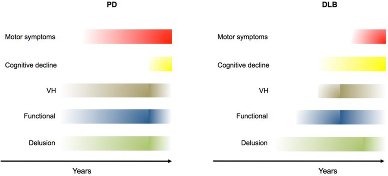
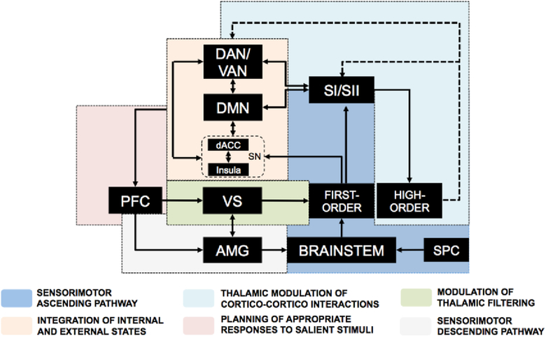
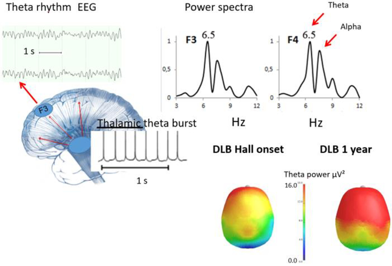
Hallucinations, delusions, and functional neurological manifestations (conversion and somatic symptom disorders) of Parkinson's disease (PD) and dementia with Lewy bodies increase in frequency with disease progression, predict the onset of cognitive decline, and eventually blend with and are concealed by dementia. These symptoms share the absence of reality constraints and can be considered comparable elements of the PD-dementia with Lewy bodies psychosis. We propose that PD-dementia with Lewy bodies psychotic disorders depend on thalamic dysfunction promoting a theta burst mode and subsequent thalamocortical dysrhythmia with focal cortical coherence to theta electroencephalogram rhythms. This theta electroencephalogram activity, also called fast-theta or pre-alpha, has been shown to predict cognitive decline and fluctuations in Parkinson's disease with dementia and dementia with Lewy bodies. These electroencephalogram alterations are now considered a predictive marker for progression to dementia. The resulting thalamocortical dysrhythmia inhibits the frontal attentional network and favors the decoupling of the default mode network. As the default mode network is involved in integration of self-referential information into conscious perception, unconstrained default mode network activity, as revealed by recent imaging studies, leads to random formation of connections that link strong autobiographical correlates to trivial stimuli, thereby producing hallucinations, delusions, and functional neurological disorders. The thalamocortical dysrhythmia default mode network decoupling hypothesis provides the rationale for the design and testing of novel therapeutic pharmacological and nonpharmacological interventions in the context of PD, PD with dementia, and dementia with Lewy bodies. © 2019 International Parkinson and Movement Disorder Society.

Keywords: Parkinson's disease; default mode network; functional disorders; hallucinations; somatic symptoms; thalamus.


Figures

Figure 1.
Figure 1.. Progression of clinical symptoms in PD and DLB.
The panel depicts a schematic representation of the time course of hallucinations, functional-somatoform disorders, delusions, motor symptoms, and dementia in PD and DLB. Color hues indicate the severity and frequency of PD-DLB-related disorders in the patient population. Abbreviations: PD: Parkinson’s disease; DLB: Dementia with Lewy Bodies; VH=visual hallucinations.
Figure 2.
Figure 2.. Network interplay involved in visual processing.
This diagram depicts the physiological interplay of networks that control visual processing. Upon physiological stimulation, the visual inputs from the retina reach the primary visual cortex through activation of the first-order thalamic nuclei. The dorsal (DAN) and ventral (VAN) networks integrate visual signals from the primary cortex and, in concert with salience network (SN), select the more significant peripheral stimuli. The DMN integrates inputs from the hippocampus and amygdala and contributes to shaping visual processing by integrating emotion- or memory-enriched introspective information. The high-order thalamic nuclei modulate cortical network activity via a negative feedback mechanism. The overall net input of these activities converges on the prefrontal cortex (PFC) that manages cognitive representation of salient stimuli. The model envisions that the activity of the SN is complementary to the DMN. The SN is a key checkpoint for anticorrelating activities of the DMN and the task-positive fronto-parietal network, thereby controlling the appropriateness of the responses given to salient stimuli. Altered engagement of the SN promotes decoupling of the DMN from the fronto-parietal network and offers a functional substrate for the onset of psychosis. Abbreviations: dorsal anterior cingulate cortex, dACC; Default Mode Network, DMN; Dorsal attention network, DAN; prefrontal cortex, PFC; hippocampus, HP; inferior temporal, IT; Ventral attention network, VAN; salience network, SN.
Figure 3.
Figure 3.. Network interplay involved in somatosensory processing.
This diagram depicts the physiological interplay of networks that control somatosensory processing. The somatosensory inputs reach the thalamus through the spinal cord (SPC) and the brainstem. The first-order thalamic nuclei then filter and transmit peripheral stimuli to the somatosensory cortices The SN participates by integrating ascending signals to somatosensory cortices through modulation of the DAN and VAN networks that, in turn, discriminate and select the more significant peripheral inputs. For its part, the DMN receives inputs from the hippocampus and amygdala and contributes to shaping sensory processing by integrating emotion- or memory-enriched introspective information. These network dynamics are regulated by activity of high-order thalamic nuclei that receive inputs from somatosensory cortices and modulate network activity via negative feedback mechanisms. The overall net input of these activities converges on the PFC that, throughout its dorsal region, plans appropriate responses to salient triggers and, via its medial portion and, in conjunction with the ventro-striatal region (VS), modulates thalamic filtering as well as the limbic response. Abbreviations: amygdala, AMG; dorsal anterior cingulate cortex, dACC; Default Mode Network, DMN; Dorsal attention network, DAN; Salience network, SN; spinal cord, SPC; somatosensory cortices, SI/SII, ventral striatum, VS.
Figure 4.
Figure 4.. EEG correlates of Theta inscription.
The top right panel depicts frequency analysis with power spectra exhibiting theta bands. The bottom left panel shows theta burst frequencies that originate in thalamic neurons to produce thalamocortical dysrhythmia (TCD). The bottom right panel shows the topography of the theta activity in a DLB patient recorded at the onset of hallucinations and one year later.

References

    1. Goetz CG, Stebbins GT, Ouyang B. Visual plus nonvisual hallucinations in Parkinson’s disease: development and evolution over 10 years. Mov Disord 2011;26(12):2196–2200. - PubMed
    1. Diederich NJ, Fenelon G, Stebbins G, Goetz CG. Hallucinations in Parkinson disease. Nat Rev Neurol 2009;5(6):331–342. - PubMed
    1. Fenelon G, Mahieux F, Huon R, Ziegler M. Hallucinations in Parkinson’s disease: prevalence, phenomenology and risk factors. Brain 2000;123 (Pt 4):733–745. - PubMed
    1. Ffytche DH, Creese B, Politis M, et al. The psychosis spectrum in Parkinson disease. Nat Rev Neurol 2017;13(2):81–95. - PMC - PubMed
    1. Goetz CG, Leurgans S, Pappert EJ, Raman R, Stemer AB. Prospective longitudinal assessment of hallucinations in Parkinson’s disease. Neurology 2001;57(11):2078–2082. - PubMed

MeSH terms