. 2019 Jul 15;15(1):245.
doi: 10.1186/s12917-019-1997-3.

Canine platelets express functional Toll-like receptor-4: lipopolysaccharide-triggered platelet activation is dependent on adenosine diphosphate and thromboxane A2 in dogs

Affiliations

Canine platelets express functional Toll-like receptor-4: lipopolysaccharide-triggered platelet activation is dependent on adenosine diphosphate and thromboxane A2 in dogs

Ronald H L Li et al. BMC Vet Res. .

Abstract

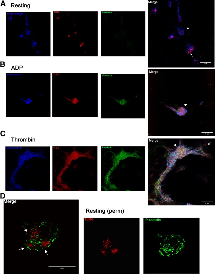
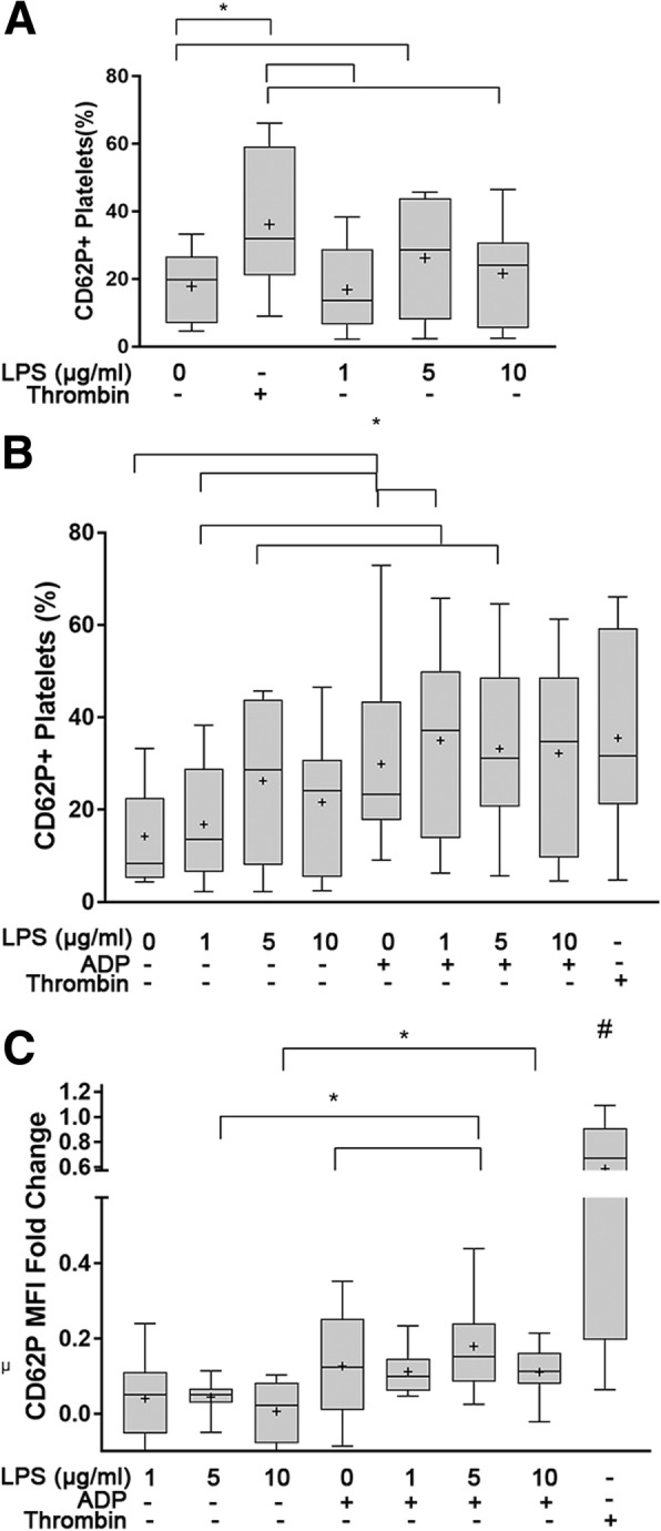
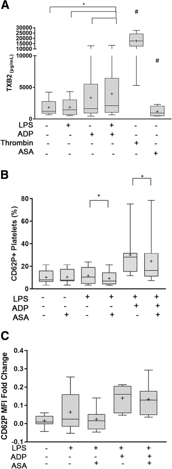
Background: Functional Toll-like receptor 4 (TLR4) has been characterized in human and murine platelets indicating that platelets play a role in inflammation and hemostasis during sepsis. It is unclear whether canine platelets could express functional TLR4 by responding to its ligand, lipopolysaccharide (LPS). We sought to determine if dogs express functional TLR4 and if LPS-induced platelet activation requires co-stimulation with ADP or thromboxane A2 (TxA2). Canine platelets were unstimulated (resting) or activated with thrombin or ADP prior to flow cytometric or microscopic analyses for TLR4 expression. We treated resting or ADP-primed platelets with LPS in the absence or presence of acetylsalicylic acid (ASA) and inhibited TLR4 with function blocking antibody or LPS from Rhodobacter sphaeroides (LPS-RS).

Results: We discovered that dog platelets have variable TLR4 expression, which was upregulated following thrombin or ADP activation. LPS augmented P-selectin expression and thromboxane B2 secretion in ADP-primed platelets via TLR4. Inhibition of cyclooxygenase by ASA attenuated LPS-mediated P-selectin expression demonstrating that TLR4 signaling in platelets is partially dependent on TxA2 pathway.

Conclusion: Expression of functional TLR4 on canine platelets may contribute to hypercoagulability in clinical septic dogs. Cyclooxygenase and TxA2 pathways in TLR4-mediated platelet activation may present novel therapeutic targets in dogs with sepsis.

Keywords: Aspirin; Inflammation; Pattern recognition receptor; Platelet-priming; Sepsis; Thrombosis.


Conflict of interest statement

The authors declare that they have no competing interests.

Figures

Fig. 1
Fig. 1
Platelet activation upregulates surface TLR4 expression. Platelet surface TLR4 expression was measured on isolated platelets from 10 dogs using flow cytometry. a,c Representative histograms of resting (unstimulated) platelets (grey) and activated platelets (clear) indicating increase in surface TLR4 expression following thrombin (a) and ADP (c) activation. b Thrombin stimulation led to significant increase in percentage (%) of TLR4-positive platelets compared to resting platelets. d Platelets stimulated with 10 μM ADP had significantly higher percentage of TLR-positive platelets. First and third quartiles were represented by the lower and upper boundaries, respectively. + and the line within the box represents the mean and median, respectively. Whiskers represent the range of data. * p < 0.05
Fig. 2
Fig. 2
Representative confocal and super resolution immunofluorescence microscopy demonstrating TLR4 expression on a resting, b ADP-, c thrombin-activated and d permabilized resting canine platelets. a-c Following activation, platelets were fixed and stained for TLR4 (red), P-selectin (green) and the integrin β3 (blue). a In the absence of platelet agonists, resting platelets as outlined by the abundant integrin β3 show limited to no expression of P-selectin on the membrane surface. Note the aggregregated appearance of exteriorized TLR4 (arrow) b,c In ADP- and thrombin-activated platelets, TLR4 expression is upregulated and is evently distributed across the membrane surface. Note the formation of pseudopodia and colocalization of TLR4 and P-selectin (arrowheads) on activated platelets c Extensive platelet aggregation can be seen in thrombin-activated platelets. Scale bar = 5 μm. Original magnification 100x d Resting platelets were fixed, permabilized and stained for TLR4 (red) and P-selectin (green). A single z-plane is shown here to demonstrate the presence of alpha-granules within a platelet (asterisk). Intracellular TLR4 can be seen within the alpha-granules (arrows). Scale bar = 4 μm. Experiment was replicated twice from platelets isolated from 2 dogs
Fig. 3
Fig. 3
Variability of P-selectin expression on unstimulated/resting platelets. Representative histograms and scatter plots of flow cytometric analysis of unstimulated platelets isolated in 2 dogs on the same day. Platelets were identified by their light scatter properties and binding of CD61. Histogram demonstrates the number of activated platelets shown as P-selectin positive. a Resting platelets with mimimal P-selectin positive platelets and characteristic light scatter profile of canine platelets. b Unstimulated platelets collected from a different dog showed increased P-selectin-positive platelets with side-scatter property indicative of increased degranulation (arrow)
Fig. 4
Fig. 4
ADP-priming augments LPS-mediated alpha-granule secretion in canine platelets. Platelet alpha-granule secretion was assessed by surface P-selectin (CD62P) measured as percent (%) positive or mean fluorescence intensity (MFI) fold change on isolated platelets from 10 dogs using flow cytometry. Thrombin-stimulated platelets served as positive control. a Platelets were treated with 0, 1, 5, or 10 μg/ml LPS. Only platelets treated with 5 μg/mL LPS had significant increase in CD62P+ platelets compared to unstimulated platelets. b,c Platelets were primed with 10 μM ADP prior to stimulation with 0, 1, 5 or 10 μg/ml LPS. LPS stimulation with 1 μg/mL LPS in ADP-primed platelets significantly elevated the percentage of CD62P+ platelets b but did not increase MFI fold change c compared to ADP-primed platelets without LPS. LPS at 5 μg/mL significantly increase CD62P MFI fold change in ADP-primed platelets relative to those without LPS. ADP priming increased platelet response to LPS at 1 (b), 5 μg/mL (b,c) and 10 μg/mL (c). First and third quartiles were represented by the lower and upper boundaries, respectively. + and the line within the box represents the mean and median, respectively. Whiskers represent the range of data. * p < 0.05, # All treatments were significantly different (p < 0.05) from positive control
Fig. 5
Fig. 5
LPS amplifies ADP-mediated thromboxane B2 (TxB2) secretion and inhibition of platelet cyclooxygenase 1 attenuates LPS-mediated alpha-granule secretion. a Platelet TxB2 concentration was measured by ELSIA from platelet supernatant in 10 dogs. In the presence or absence of ADP, platelets were treated with 5 μg/ml LPS. Thrombin-stimulated and acetyl salicylic acid (ASA)-treated platelets served as positive and negative controls, respectively. LPS-treated platelets did not augment TxB2 production compared to unstimulated platelets and ADP-treated platelets. ADP priming in LPS-treated platelets led to more TxB2 than ADP-treated platelets, LPS-treated platelets and unstimulated platelets. b,c Isolated platelets from 10 dogs were pretreated with 100 μM acetylsalicyclic acid (ASA) prior to treatment with 5 μg/ml LPS with or without ADP. Platelet alpha-granule secretion was assessed by P-selectin (CD62P) measured as percent (%) positive b or mean fluorescence intensity (MFI) fold change c using flow cytometry. ASA significantly decreased the % P-selectin positive platelets a but not P-selectin MFI fold change b in LPS-treated platelets in the absence or presence of ADP-priming. First and third quartiles were represented by the lower and upper boundaries, respectively, and the line within the box represents the median. + represents the mean. Whiskers represent the range of date. * p < 0.05, # Significance between all treatments and controls (p<0.05)
Fig. 6
Fig. 6
LPS-mediated alpha-granule secretion is dependent on platelet TLR4. Platelet P-selectin (CD62P) measured as percent (%) positive platelets or mean fluorescence intensity (MFI) fold change on isolated platelets (1 × 107/ml) from 10 dogs using flow cytometry. a, b Platelets were treated with either 50 μg/ml TLR4 function blocking antibody or 50 μg/ml IgG2a before LPS stimulation (5 μg/ml) in absence or presence of ADP. TLR4 inhibition in LPS-treated had minimal effect on attenuating the numbers of P-selectin positive platelets (a) but significantly decreased P-selectin MFI fold change b in the presence or absence of ADP priming. Pretreatment of platelets with LPS-RS before LPS stimulation significantly decreased number of P-selectin postive platelets and MFI fold change only in ADP-primed platelets. First and third quartiles were represented by the lower and upper boundaries, respectively, and the line within the box represents the median. Whiskers represent the range of data. + represents the mean * p < 0.05, #Significance between all treatments and isotype controls (p<0.05)
Fig. 7
Fig. 7
Schematic diagram of LPS-mediated platelet activation and TLR4 expression in canine platelets. ADP activation via P2Y1 or P2Y12 receptor upregulates surface TLR4 expression. TLR4 trafficking to cell membrane from granules may be mediated by alpha-granule secretion. LPS binding protein (LBP) presents LPS to CD14 forming a heterodimeric complex with TLR4 and myeloid differentiation protein 2 (MD-2). Downstream signaling pathway of TLR4 leads to α-granule secretion, which is amplified by ADP and thromboxane A2 (TxA2). Activation of G-protein coupled receptors, P2Y1/P2Y12 and thromboxane receptor (TP), leads to phospholipase C (PLC) activation and, subsequently, 1,4,5-triphosphate (IP3) and diacylglycerol (DAG) for intracellular calcium release and alpha-granule secretion. LPS acts synergistically with ADP to increase generation of TxA2, serving a positive feedback mediator. TLR4 and ADP signaling activates cyclooxygenase-1 (COX-1), which converts arachidonic acid (AA) to TxA2, likely by the Akt/p38 MAPK pathway

Similar articles

Cited by

References

    1. Dickinson AE, Summers JF, Wignal J, Boag AK, Keir I. Impact of appropriate empirical antimicrobial therapy on outcome of dogs with septic peritonitis. J Vet Emerg Crit Care (San Antonio) 2015;25(1):152–159. doi: 10.1111/vec.12273. - DOI - PubMed
    1. Wong C, Epstein SE, Westropp JL. Antimicrobial susceptibility patterns in urinary tract infections in dogs (2010-2013) J Vet Intern Med. 2015;29(4):1045–1052. doi: 10.1111/jvim.13571. - DOI - PMC - PubMed
    1. Proulx A, Hume DZ, Drobatz KJ, Reineke EL. In vitro bacterial isolate susceptibility to empirically selected antimicrobials in 111 dogs with bacterial pneumonia. J Vet Emerg Crit Care (San Antonio) 2014;24(2):194–200. doi: 10.1111/vec.12128. - DOI - PubMed
    1. Duerschmied D, Bode C, Ahrens I. Immune functions of platelets. Thromb Haemost. 2014;112(4):678–691. - PubMed
    1. Watson CN, Kerrigan SW, Cox D, Henderson IR, Watson SP, Arman M. Human platelet activation by Escherichia coli: roles for FcgammaRIIA and integrin alphaIIbbeta3. Platelets. 2016;27(6):535–540. doi: 10.3109/09537104.2016.1148129. - DOI - PMC - PubMed

MeSH terms