

Imaging-based clusters in former smokers of the COPD cohort associate with clinical characteristics: the SubPopulations and intermediate outcome measures in COPD study (SPIROMICS)
- PMID: 31307479
- PMCID: PMC6631615
- DOI: 10.1186/s12931-019-1121-z
Imaging-based clusters in former smokers of the COPD cohort associate with clinical characteristics: the SubPopulations and intermediate outcome measures in COPD study (SPIROMICS)
Abstract
Background: Quantitative computed tomographic (QCT) imaging-based metrics enable to quantify smoking induced disease alterations and to identify imaging-based clusters for current smokers. We aimed to derive clinically meaningful sub-groups of former smokers using dimensional reduction and clustering methods to develop a new way of COPD phenotyping.
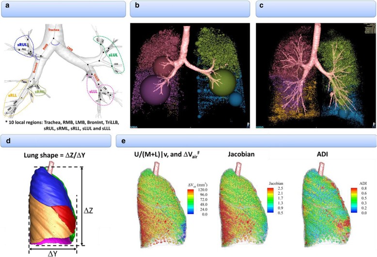
Methods: An imaging-based cluster analysis was performed for 406 former smokers with a comprehensive set of imaging metrics including 75 imaging-based metrics. They consisted of structural and functional variables at 10 segmental and 5 lobar locations. The structural variables included lung shape, branching angle, airway-circularity, airway-wall-thickness, airway diameter; the functional variables included regional ventilation, emphysema percentage, functional small airway disease percentage, Jacobian (volume change), anisotropic deformation index (directional preference in volume change), and tissue fractions at inspiration and expiration.
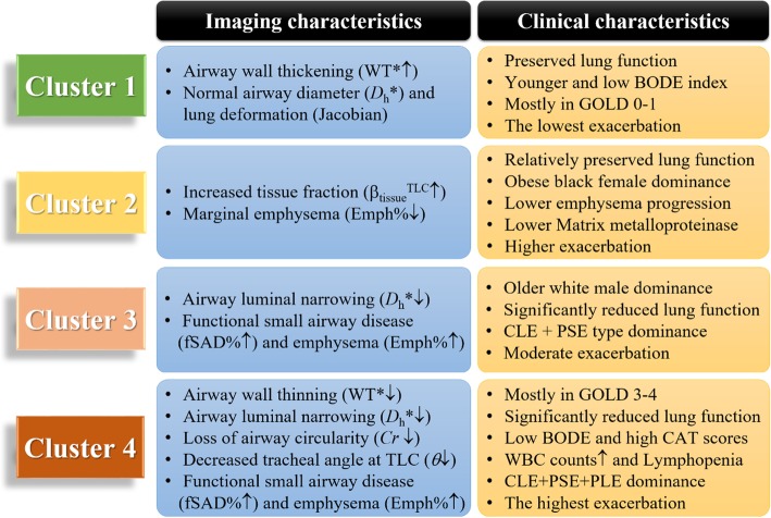
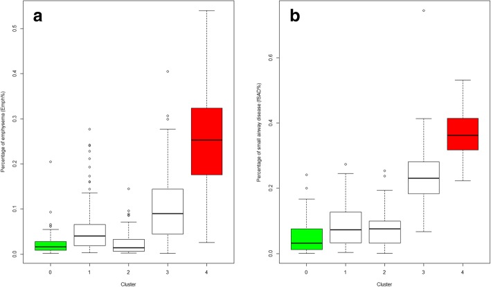
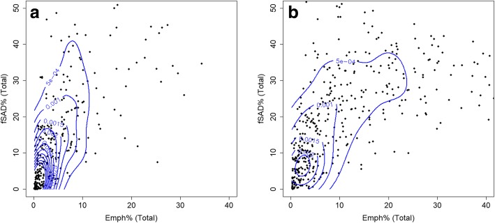
Results: We derived four distinct imaging-based clusters as possible phenotypes with the sizes of 100, 80, 141, and 85, respectively. Cluster 1 subjects were asymptomatic and showed relatively normal airway structure and lung function except airway wall thickening and moderate emphysema. Cluster 2 subjects populated with obese females showed an increase of tissue fraction at inspiration, minimal emphysema, and the lowest progression rate of emphysema. Cluster 3 subjects populated with older males showed small airway narrowing and a decreased tissue fraction at expiration, both indicating air-trapping. Cluster 4 subjects populated with lean males were likely to be severe COPD subjects showing the highest progression rate of emphysema.
Conclusions: QCT imaging-based metrics for former smokers allow for the derivation of statistically stable clusters associated with unique clinical characteristics. This approach helps better categorization of COPD sub-populations; suggesting possible quantitative structural and functional phenotypes.
Keywords: COPD; Emphysema; Former smokers; Functional small airway disease; Imaging-based cluster analysis.
Conflict of interest statement
The authors declare that they have no competing interests.
Figures
Similar articles
-
Longitudinal Imaging-Based Clusters in Former Smokers of the COPD Cohort Associate with Clinical Characteristics: The SubPopulations and Intermediate Outcome Measures in COPD Study (SPIROMICS).Int J Chron Obstruct Pulmon Dis. 2021 May 31;16:1477-1496. doi: 10.2147/COPD.S301466. eCollection 2021. Int J Chron Obstruct Pulmon Dis. 2021. PMID: 34103907 Free PMC article.
-
Imaging-based clusters in current smokers of the COPD cohort associate with clinical characteristics: the SubPopulations and Intermediate Outcome Measures in COPD Study (SPIROMICS).Respir Res. 2018 Sep 18;19(1):178. doi: 10.1186/s12931-018-0888-7. Respir Res. 2018. PMID: 30227877 Free PMC article.
-
Occupational Exposures and Computed Tomographic Imaging Characteristics in the SPIROMICS Cohort.Ann Am Thorac Soc. 2018 Dec;15(12):1411-1419. doi: 10.1513/AnnalsATS.201802-150OC. Ann Am Thorac Soc. 2018. PMID: 30339479 Free PMC article.
-
Quantitative computed tomography measurements to evaluate airway disease in chronic obstructive pulmonary disease: Relationship to physiological measurements, clinical index and visual assessment of airway disease.Eur J Radiol. 2016 Nov;85(11):2144-2151. doi: 10.1016/j.ejrad.2016.09.010. Epub 2016 Sep 13. Eur J Radiol. 2016. PMID: 27776670 Free PMC article. Review.
-
Imaging Advances in Chronic Obstructive Pulmonary Disease. Insights from the Genetic Epidemiology of Chronic Obstructive Pulmonary Disease (COPDGene) Study.Am J Respir Crit Care Med. 2019 Feb 1;199(3):286-301. doi: 10.1164/rccm.201807-1351SO. Am J Respir Crit Care Med. 2019. PMID: 30304637 Free PMC article. Review.
Cited by
-
Contribution of adaptive immunity to human COPD and experimental models of emphysema.Physiol Rev. 2023 Apr 1;103(2):1059-1093. doi: 10.1152/physrev.00036.2021. Epub 2022 Oct 6. Physiol Rev. 2023. PMID: 36201635 Free PMC article. Review.
-
Radiomic phenotyping of the lung parenchyma in a lung cancer screening cohort.Sci Rep. 2023 Feb 4;13(1):2040. doi: 10.1038/s41598-023-29058-1. Sci Rep. 2023. PMID: 36739358 Free PMC article.
-
A titratable murine model of progressive emphysema using tracheal porcine pancreatic elastase.Sci Rep. 2023 Sep 14;13(1):15259. doi: 10.1038/s41598-023-41527-1. Sci Rep. 2023. PMID: 37709810 Free PMC article.
-
Latent traits of lung tissue patterns in former smokers derived by dual channel deep learning in computed tomography images.Sci Rep. 2021 Mar 1;11(1):4916. doi: 10.1038/s41598-021-84547-5. Sci Rep. 2021. PMID: 33649381 Free PMC article.
-
Artificial intelligence in thoracic imaging: the transition from research to practice.Eur Radiol. 2023 Sep;33(9):6318-6319. doi: 10.1007/s00330-023-09635-8. Epub 2023 Apr 25. Eur Radiol. 2023. PMID: 37186215 No abstract available.
References
-
- Miniño AM, Murphy SL, Xu J, Kochanek KD. Deaths: final data for 2008. Natl Vital Stat Rep Cent Dis Control Prev Natl Cent Health Stat Natl Vital Stat Syst. 2011;59:1–126. - PubMed
-
- Couper D, LaVange LM, Han M, Barr RG, Bleecker E, Hoffman EA, Kanner R, Kleerup E, Martinez FJ, Woodruff PG, Rennard S, SPIROMICS Research Group Design of the Subpopulations and Intermediate Outcomes in COPD study (SPIROMICS) Thorax. 2014;69:491–494. doi: 10.1136/thoraxjnl-2013-203897. - DOI - PMC - PubMed
-
- Agusti A, Calverley PMA, Celli B, Coxson HO, Edwards LD, Lomas DA, MacNee W, Miller BE, Rennard S, Silverman EK, Tal-Singer R, Wouters E, Yates JC, Vestbo J. Evaluation of COPD longitudinally to identify predictive surrogate endpoints (ECLIPSE) investigators. Characterisation of COPD heterogeneity in the ECLIPSE cohort. Respir Res. 2010;11:122. doi: 10.1186/1465-9921-11-122. - DOI - PMC - PubMed
-
- Sieren JP, Newell JD, Barr RG, Bleecker ER, Burnette N, Carretta EE, Couper D, Goldin J, Guo J, Han MK, Hansel NN, Kanner RE, Kazerooni EA, Martinez FJ, Rennard S, Woodruff PG, Hoffman EA. SPIROMICS protocol for multicenter quantitative computed tomography to phenotype the lungs. Am J Respir Crit Care Med. 2016;194:794–806. doi: 10.1164/rccm.201506-1208PP. - DOI - PMC - PubMed
-
- Choi S, Hoffman EA, Wenzel SE, Castro M, Fain S, Jarjour N, Schiebler ML, Chen K, Lin C-L, Heart N. Lung and blood Institute’s severe asthma research program. Quantitative computed tomographic imaging-based clustering differentiates asthmatic subgroups with distinctive clinical phenotypes. J Allergy Clin Immunol. 2017;140:690–700.e8. doi: 10.1016/j.jaci.2016.11.053. - DOI - PMC - PubMed
MeSH terms
Grants and funding
- U24 HL141762/HL/NHLBI NIH HHS/United States
- S10 OD018526/OD/NIH HHS/United States
- P30 CA086862/CA/NCI NIH HHS/United States
- K24 HL137013/HL/NHLBI NIH HHS/United States
- U01 HL114494/HL/NHLBI NIH HHS/United States
- U01 HL137880/HL/NHLBI NIH HHS/United States
- R01 HL130506/HL/NHLBI NIH HHS/United States
- S10 RR022421/RR/NCRR NIH HHS/United States
- R01 HL112986/HL/NHLBI NIH HHS/United States
- P30 ES005605/ES/NIEHS NIH HHS/United States
- NIH grants U01-HL114494, R01-HL112986 and S10-RR022421/NH/NIH HHS/United States
- P30 ES006694/ES/NIEHS NIH HHS/United States
LinkOut - more resources
Full Text Sources
Medical
Molecular Biology Databases
