

Truncated Rep protein of porcine circovirus 2 (PCV2) caused by a naturally occurring mutation reduced virus replication in PK15 cells
- PMID: 31307486
- PMCID: PMC6632220
- DOI: 10.1186/s12917-019-1984-8
Truncated Rep protein of porcine circovirus 2 (PCV2) caused by a naturally occurring mutation reduced virus replication in PK15 cells
Abstract
Background: Porcine circovirus 2 (PCV2) is the causative agent of porcine circovirus-associated diseases (PCVADs). The infection of PCV2 is widespread and has serious consequence, thereby causing significant economic losses in the swine industry worldwide. Previously, we found that a strain named YiY-3-2-3 has a naturally occurring point mutation (G710 to A710) in ORF1 region, which leads to a shorten product of the rep gene (945 to 660 base pair). Importantly, the Rep protein is responsible for genome replication of PCV2. To explore the effects of this mutation on the PCV2 replication, in the current study we constructed infectious clone of this IF-YiY-3-2-3, as well as those of its two parental strains of IF-YiY-3-2-1 and IF-YiY-3-2-10. Subsequently, these infectious clones which have 1.1 copy of PCV2 genome of their corresponding strains were transfected into PK15 cells to obtain rescued viruses, respectively.
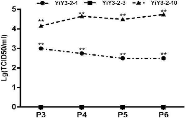
Results: Though all of the three infectious clones could be rescued, the copy number and infectivity of these rescued viruses were significantly different, as analyzed by fluorescence quantitative PCR, Tissue culture infectious dose 50 (TCID50), and indirect immunofluorescence assay (IFA). Notably, whether the PCV2 copy number, viral titer or the infectivity of rescued viruses from infectious clone IF-YiY-3-2-3 was significantly less than those of its parental clones. Meanwhile, the spatial structure of the Rep protein from the IF-YiY-3-2-3 displayed an apparent truncation at the C-terminal.
Conclusions: These findings therefore suggest that the Rep protein with truncated C-terminal would reduce virus replication and infectivity, and there might also exist both favorable and unfavorable mutations in the ORF1 of PCV2 in the process of its evolution.
Keywords: Infectious clone; PCV2 evolution; Porcine circovirus 2(PCV2); Rescued viruses; Truncated Rep protein; Virus replication.
Conflict of interest statement
The authors declare that they have no competing interests.
Figures
Similar articles
-
Mutation of the sixth amino acid of the Rep protein has no effect on porcine circovirus 2b but enhances porcine circovirus 2d replication in vitro.Arch Virol. 2021 Nov;166(11):3189-3192. doi: 10.1007/s00705-021-05217-5. Epub 2021 Sep 15. Arch Virol. 2021. PMID: 34524537
-
The correlation between the mutual deletions of amino acids within porcine circovirus rep protein and the discrepancy of replication.Microb Pathog. 2018 Apr;117:327-334. doi: 10.1016/j.micpath.2018.02.045. Epub 2018 Feb 26. Microb Pathog. 2018. PMID: 29496526
-
[Biological character of porcine circovirus type 2 with site mutation at interferon stimulation reaction element (ISRE) like region in Rep gene promoter].Wei Sheng Wu Xue Bao. 2009 Sep;49(9):1217-22. Wei Sheng Wu Xue Bao. 2009. PMID: 20030061 Chinese.
-
Current understanding of genomic DNA of porcine circovirus type 2.Virus Genes. 2014 Aug;49(1):1-10. doi: 10.1007/s11262-014-1099-z. Epub 2014 Jul 11. Virus Genes. 2014. PMID: 25011695 Review.
-
[Porcine circovirus type 2 and PCV2-systemic disease--a review].Sheng Wu Gong Cheng Xue Bao. 2015 Jun;31(6):880-91. Sheng Wu Gong Cheng Xue Bao. 2015. PMID: 26672364 Review. Chinese.
Cited by
-
Humoral and Cellular Immune Responses Induced by Bivalent DNA Vaccines Expressing Fusion Capsid Proteins of Porcine Circovirus Genotypes 2a and 2b.Vaccines (Basel). 2024 Mar 18;12(3):324. doi: 10.3390/vaccines12030324. Vaccines (Basel). 2024. PMID: 38543958 Free PMC article.
-
Serum investigation of antibodies against porcine circovirus 4 Rep and Cap protein in Jiangxi Province, China.Front Microbiol. 2022 Oct 21;13:944679. doi: 10.3389/fmicb.2022.944679. eCollection 2022. Front Microbiol. 2022. PMID: 36338086 Free PMC article.
-
Mutation of the sixth amino acid of the Rep protein has no effect on porcine circovirus 2b but enhances porcine circovirus 2d replication in vitro.Arch Virol. 2021 Nov;166(11):3189-3192. doi: 10.1007/s00705-021-05217-5. Epub 2021 Sep 15. Arch Virol. 2021. PMID: 34524537
-
Retinol binding protein 4 restricts PCV2 replication via selective autophagy degradation of viral ORF1 protein.Commun Biol. 2024 Nov 5;7(1):1438. doi: 10.1038/s42003-024-07052-1. Commun Biol. 2024. PMID: 39500783 Free PMC article.
-
Development and Primary Application of an Indirect ELISA Based on Rep Protein to Analyze Antibodies against Porcine Cocirvirus-like Virus (PCLV).Viruses. 2022 Jun 27;14(7):1398. doi: 10.3390/v14071398. Viruses. 2022. PMID: 35891379 Free PMC article.
References
-
- Allan GM, Ellis JA: Porcine circoviruses: a review. Journal of veterinary diagnostic investigation : official publication of the American Association of Veterinary Laboratory Diagnosticians, Inc 2000, 12(1):3–14. - PubMed
-
- Grau-Roma L, Crisci E, Sibila M, Lopez-Soria S, Nofrarias M, Cortey M, Fraile L, Olvera A, Segales J. A proposal on porcine circovirus type 2 (PCV2) genotype definition and their relation with postweaning multisystemic wasting syndrome (PMWS) occurrence. Vet Microbiol. 2008;128(1–2):23–35. doi: 10.1016/j.vetmic.2007.09.007. - DOI - PubMed
MeSH terms
Substances
Grants and funding
LinkOut - more resources
Full Text Sources
Miscellaneous
