. 2019 Jul 15;38(1):308.
doi: 10.1186/s13046-019-1295-8.

Synergistic killing effects of homoharringtonine and arsenic trioxide on acute myeloid leukemia stem cells and the underlying mechanisms

Affiliations

Synergistic killing effects of homoharringtonine and arsenic trioxide on acute myeloid leukemia stem cells and the underlying mechanisms

Ming Tan et al. J Exp Clin Cancer Res. .

Erratum in

Abstract

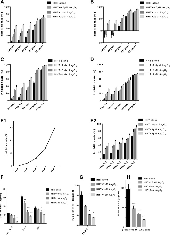
Background: At present, it is generally believed that leukemia stem cells are the source of AML, so the killing of leukemia stem cells has become important. Previous studies have suggested that HHT combined with ATO can synergistically kill U937 cells, and HHT has also demonstrated the ability to kill leukemia stem cells. We evaluated whether HHT combined with ATO can systematically kill leukemia stem cells (LSCs) and explored the synergistic effect and molecular mechanism.

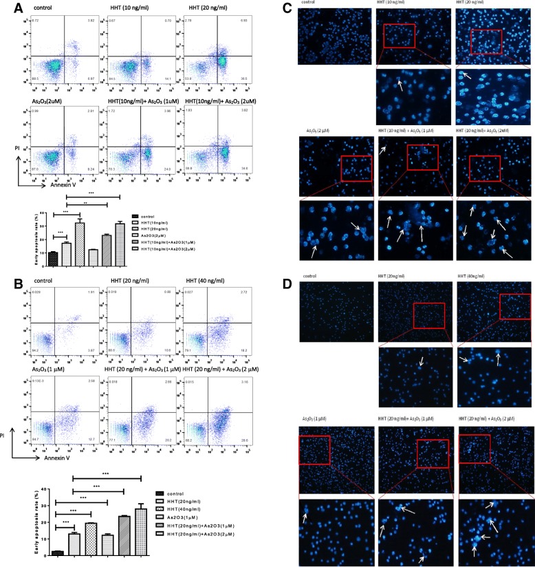
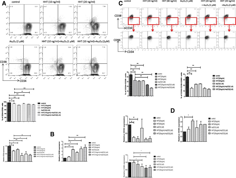
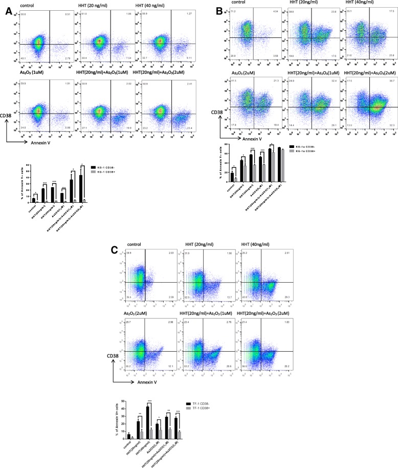
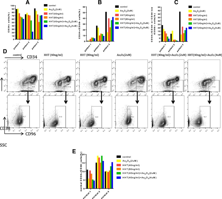
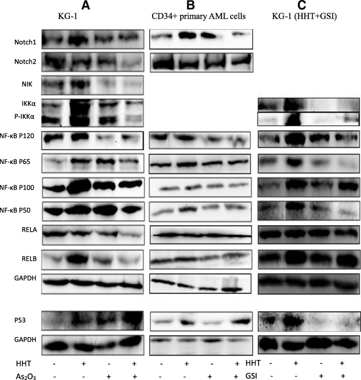
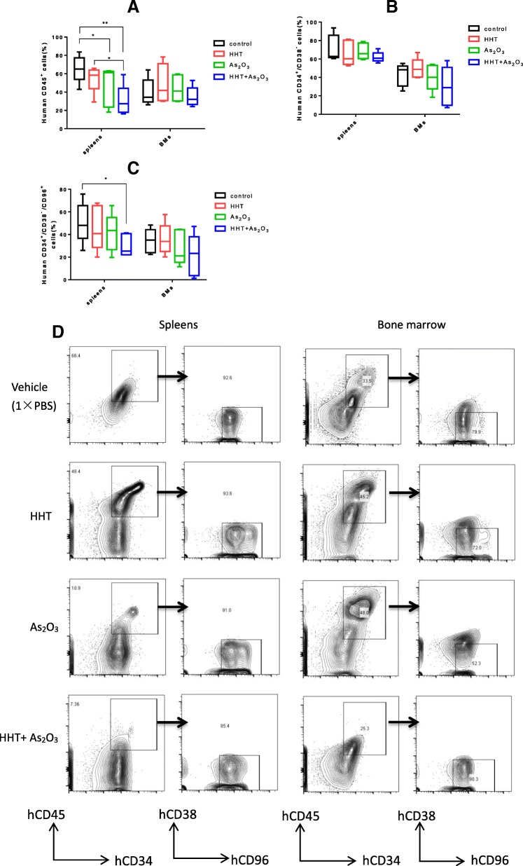
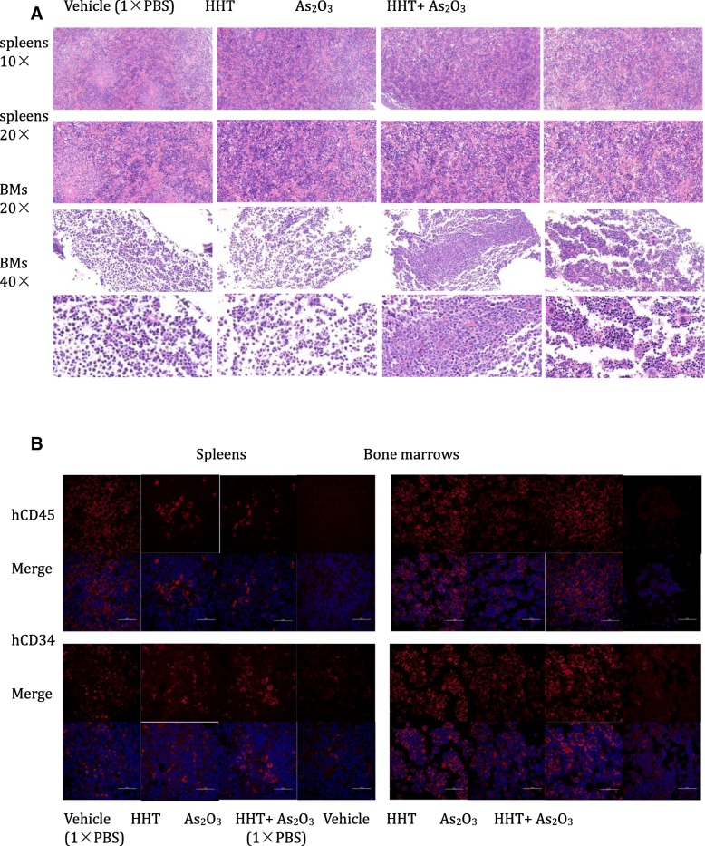
Methods: CCK-8 was used to detect cell viability. The changes of cell cycle (PI staining), apoptosis (Annexin V/PI) and surface markers (CD34, CD38, CD96, CD45) were detected by flow cytometry. The cells of CD34+ primary leukemia and CD38- KG-1, and TF-1 were separated by flow cytometry. High-throughput mRNA sequencing was used to analysis mRNA level changes after the application of the two drugs. Western blot was used to verify the changes of pathway protein expression. NRG mice were used as the receptor of xenograft model. Histological H&E staining assess the invaded ability of leukemia cells, and laser scanning confocal microscopy evaluated the molecule markers change.

Results: HHT and ATO synergistically killed KG-1 (CD34+/CD96+/CD38+/-) and Kasumi-1 (CD34+/CD38-) cells. Their combination had a stronger effect of inducing apoptosis and blocking the cell cycle than HHT or ATO administrator alone, meanwhile significantly reducing the numbers of LSCs. Further, CD34+CD38- cells in KG-1, KG-1a, TF-1, and primary leukemia cells were more sensitive to HHT and ATO. High-throughput mRNA sequencing suggested that HHT alone could significantly upregulate molecules related to the Notch, P53, and NF-κB signaling pathways. When combined with ATO, HHT further upregulated P53, whereas HHT-induced NF-κB pathway activation was significantly suppressed. Western blot analysis verified the change of protein expression in the above pathways and further demonstrated that GSI, could eliminate these effects. In vivo, HHT combined with ATO significantly reduced the LSC burden, and weakened the expression of LSC markers.

Conclusions: This is the first evidence that HHT combined with arsenic can synergistically kill LSCs in vitro and in vivo, along with identification of the underlying mechanism, highlighting a potentially effective treatment strategy.

Keywords: Arsenic trioxide; Homoharringtonine; LSC; Notch; Xenograft leukemia model.


Conflict of interest statement

The authors declare that they have no competing interests.

Figures

Fig. 1
Fig. 1
Arsenic trioxide (ATO) enhances the cytotoxic effect of homoharringtonine (HHT) in acute myeloid leukemia (AML) cell lines and primary CD34+ AML cells, and HHT and ATO synergistically damage leukemia stem-like cell lines and CD34+ primary cells (n = 3). Kasumi-1 cells (a), KG-1 cells (b), THP-1 cells (c), HEL cells (d), and primary CD34+ AML cells (e1, e2) were treated with HHT, ATO, or HHT + ATO for 2 days, and cell viability was measured by the CCK-8 assay. IC50 of HHT is calculated by using SPSS 22 (f,g,h). Error bars represent standard deviations of three independent experiments. P < 0.05*, P < 0.01**, P < 0.001***
Fig. 2
Fig. 2
Arsenic trioxide (ATO) cooperates with homoharringtonine (HHT) to induce apoptosis in leukemia stem-like cell lines. Cells were treated with HHT, ATO, or HHT + ATO for 2 days, and apoptosis of Kasumi-1 (a) and KG-1 (b) cells was detected with PI/Annexin V and FACS. Error bars represent three independent experiments. P < 0.05*, P < 0.01**, P < 0.001***. Hochest dye was used to evaluate the shape of the nuclei of Kasumi-1 (c) and KG-1 (d) cells under fluorescence microscopy
Fig. 3
Fig. 3
Arsenic trioxide (ATO) promotes the ability of homoharringtonine (HHT) to decrease the proportion of CD34+ CD38 cells. Cells were treated with HHT, ATO, or HHT + ATO for 2 days, and cell surface antigen of Kasumi-1 (a) and KG-1 (c) cells was detected using FACS. The relative expression levels of CD34, CD38, and CD96 mRNA of Kasumi-1 (b) and KG-1 (d) cells were quantified using qPCR. Error bars represent three independent experiments. P < 0.05*, P < 0.01**, P < 0.001***
Fig. 4
Fig. 4
Homoharringtonine (HHT) combined with arsenic trioxide (ATO) more effectively killed the CD34+ CD38 leukemia stem cells. Cells were treated with HHT, ATO, or HHT + ATO for 2 days, and then stained with CD38 antibody and Annexin V; the apoptosis rate of cells with different expression levels of CD38 was detected using FACS. Error bars represent three independent experiments. P < 0.05*, P < 0.01**, P < 0.001***
Fig. 5
Fig. 5
Homoharringtonine (HHT) combined with arsenic trioxide (ATO) decrease the proportion of primary leukemia stem cells (LSCs) in serum free medium with cytokine cocktail (Flt3L, SCF, IL-3 and IL-6). Quantification of frequencies of CD34+cells (a), CD34+/CD38 cells (b) and CD34+/CD38/CD96+ cells (c). (d) Display of flow cytometric analysis on bone marrow sample of patient no. 2 after treatment with HHT and ATO alone or combined. (e) Represents the proportion of normal primary CD34+/CD38- (n = 3)
Fig. 6
Fig. 6
Western bloting analysis for protein expression. KG-1 (a) and CD34+ primary cells (b) were treated with HHT, arsenic trioxide (ATO) alone or in combination for 24 h, and total proteins were separated for western blotting. (c) GSI (1µM) eliminated the influence of homoharringtonine HHT to Notch, NF-κB, and P53
Fig. 7
Fig. 7
Homoharringtonine (HHT) combined with arsenic trioxide (ATO) efficaciously eliminated the leukemia stem cells (LSCs) in a xenograft model of NRG mice (n = 5). (a) Evaluation of enrichment of KG-1 cells in the spleens and bone marrow of mice by analysis of hCD45 expression using flow cytometry. (b, c) Detection the residual of LSCs (CD34+CD38CD96+) in the spleens and bone marrow using flow cytometry. (d) Representative flow cytometric analysis of stem LSCs markers (CD34, CD38, CD96). P < 0.05*, P < 0.01**, P < 0.001***
Fig. 8
Fig. 8
Homoharringtonine (HHT) combined with arsenic trioxide (ATO) remarkably obliterated the histological infiltration of leukemia stem cells (LSCs). (a) H&E-stained sections of representative 4% paraformaldehyde-fixed spleens and bone marrow from NRG mice. (b) hCD45 and hCD34 levels were detected in the different groups by confocal laser-scanning microscopy in representative 4% paraformaldehyde-fixed spleens and bone marrow samples from NRG mice. Scale bars: 50 μm

Similar articles

Cited by

References

    1. Hope KJ, Jin L, Dick JE. Acute myeloid leukemia originates from a hierarchy of leukemic stem cell classes that differ in self-renewal capacity. Nat Immunol. 2004;5:738–743. doi: 10.1038/ni1080. - DOI - PubMed
    1. Zhang Y, Xia F, Liu X, Yu Z, Xie L, Liu L, et al. JAM3 maintains leukemia-initiating cell self-renewal through LRP5/AKT/beta-catenin/CCND1 signaling. J Clin Invest. 2018;128:1737–1751. doi: 10.1172/JCI93198. - DOI - PMC - PubMed
    1. Testa U. Leukemia stem cells. Ann Hematol. 2011;90:245–271. doi: 10.1007/s00277-010-1118-7. - DOI - PubMed
    1. Hosen N, Park CY, Tatsumi N, Oji Y, Sugiyama H, Gramatzki M, et al. CD96 is a leukemic stem cell-specific marker in human acute myeloid leukemia. Proc Natl Acad Sci U S A. 2007;104:11008–11013. doi: 10.1073/pnas.0704271104. - DOI - PMC - PubMed
    1. Liu Y, Chen F, Wang S, Guo X, Shi P, Wang W, et al. Low-dose triptolide in combination with idarubicin induces apoptosis in AML leukemic stem-like KG1a cell line by modulation of the intrinsic and extrinsic factors. Cell Death Dis. 2013;4:e948. doi: 10.1038/cddis.2013.467. - DOI - PMC - PubMed

MeSH terms