. 2019 Aug 6;117(3):464-478.
doi: 10.1016/j.bpj.2019.06.013. Epub 2019 Jul 2.

Sustained Oscillations of Epithelial Cell Sheets

Affiliations

Sustained Oscillations of Epithelial Cell Sheets

Grégoire Peyret et al. Biophys J. .

Abstract

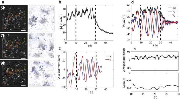
Morphological changes during development, tissue repair, and disease largely rely on coordinated cell movements and are controlled by the tissue environment. Epithelial cell sheets are often subjected to large-scale deformation during tissue formation. The active mechanical environment in which epithelial cells operate have the ability to promote collective oscillations, but how these cellular movements are generated and relate to collective migration remains unclear. Here, combining in vitro experiments and computational modeling, we describe a form of collective oscillations in confined epithelial tissues in which the oscillatory motion is the dominant contribution to the cellular movements. We show that epithelial cells exhibit large-scale coherent oscillations when constrained within micropatterns of varying shapes and sizes and that their period and amplitude are set by the smallest confinement dimension. Using molecular perturbations, we then demonstrate that force transmission at cell-cell junctions and its coupling to cell polarity are pivotal for the generation of these collective movements. We find that the resulting tissue deformations are sufficient to trigger osillatory mechanotransduction of YAP within cells, potentially affecting a wide range of cellular processes.


Figures

Figure 1
Figure 1
Coordinated oscillations of a confined epithelium. (a) Left column shows snapshots of a confluent HaCaT layer on a square pattern at various times and representative trajectories of single cells within the pattern. Right shows velocity fields from PIV measurement at the corresponding times. The scale bar represents 100 μm. (b) Temporal evolution of the average velocity magnitude. The velocity increases slowly at first and then plateaus for almost 20 h until it finally decreases. Vertical lines denote the period of time chosen to best highlight the oscillations. (c) Displacement of a single cell in the center of the domain. (d) Evolution of the two projected components—Vx=vxROI and Vy=vyROI computed on a cropped area in the center of the square—and the norm of the velocity V=(Vx2+Vy2)1/2. (e) Angular velocity of the average direction of the velocity (top) and phase shift between Vx and Vy (bottom) within the time interval shown in (b)–(d). To see this figure in color, go online.
Figure 2
Figure 2
Geometry-dependent coordinated oscillations. (a and b) Amplitude A and period T of the oscillations as a function of the size of the square confinement. (c) A snapshot of rectangular confinement of size L × W = 3500 × 500 μm is shown. The frame represents the ROI used in (d), (e), and (h). The scale bar represents 200 μm. (d) A spatiotemporal map of Vx=vxROI; velocity along the long axis x shows patches of correlated motion with a coherence size of about 500 μm. The velocity component Vx was averaged along the y direction (short axis) in the area outlined in orange in (c) and then plotted against time t and x (the long axis of the frame). (e) A spatiotemporal map of Vy=vyROI, velocity along the short axis, averaged over y and plotted against t and x, is given. (f and g) Amplitude A and period T of the oscillations in squares and rectangles as a function of the smallest dimension of the confinements. (h) Phase coherence length Lϕ as a function of the width of the rectangular pattern. Lϕ is defined as the distance over which Vx(x, t) reverses direction. All plots, mean (SD) from n3 experiments. n.s., not significant, p < 0.05, ∗∗p < 0.01, ∗∗∗p < 0.001 from a two-sample Kolmogorov-Smirnov test. To see this figure in color, go online.
Figure 3
Figure 3
Oscillation-induced cell contractions lead to YAP translocation and actin reorganization. (a) Example cells expressing histone-GFP followed in time by live imaging. Scale bar is 100 μm. (b) Corresponding cell-density map computed by counting the nuclei in 128 × 128 pixels windows overlapping by 87.5%. (c) A spatiotemporal kymograph of the divergence of the velocity in a square of width W = 500 μm, showing alternating phases of compression and expansion. (d) Immunostaining of YAP on a fixed sample of confined HaCaT cells with zoomed areas (marked with a square) illustrating cytoplasmic (left) and nuclear (right) YAP localization. (e) Temporal evolution of YAP-NCR (squares) and cell area (circles) for an example cell. The YAP-NCR was defined as the mean YAP-GFP fluorescence intensity in the nucleus divided by the mean YAP-GFP fluorescence intensity in the cytoplasm. The event of spreading at t ∼ 300 min corresponds to a sudden increase in YAP-NCR. (f) Time cross correlation of the single cell area and YAP-NCR, averaged over the cell population in three independent experiments. Standard error of the mean (SEM) is for n = 15 cells. (g) YAP-NCR as a function of the cell area in fixed samples of confined epithelia. (h) Confocal picture of actin staining in the basal plane of fixed HaCaT cells confined on a square of width W = 500 μm, overlaid with the local cell density. The scale bar represents 100 μm. Right displays zoomed areas showing different organizations of actin fibers with respect to the local density (1,3, low density; 2,4, high density). The scale bar represents 20 μm. The four pictures have the same scale and contrast. To see this figure in color, go online.
Figure 4
Figure 4
Traction force and cell polarity are coupled to cell motion. (a) A map of the magnitude of the traction forces. The scale bar represents 100 μm. (b) A map of the pressure P = −Tr(σ)/2 within the confined tissue. The scale bar represents 100 μm. (c) Confocal images of actin staining in fixed HaCaT cells fixed during the oscillation phase. The arrows denote cell-cell junctions under tension. The scale bar represents 20 μm (same scale for three pictures). (d) The averages of vx and −Tx in a central square ROI, projected on the x axis, oscillate with similar period but are out of phase. (e) Normalized cross correlation between v and −T as a function of the time lag Δt. The correlation has a marked peak at Δt = 0.5 h for both W = 200 and 500 μm. Mean (SD) is of n = 11 and 8, respectively. (f) Top shows a schematic of the cell polarity: the orientation of the lamellipodium (red) with respect to the cell body (turquoise) defines the polarity (vector p), which can be in a different direction from the velocity v. The traction force T is a linear combination of the two. Bottom shows an example of a reorientation event (fluorescent cell embedded in a nonfluorescent confluent monolayer). F-actin in the basal plane (z = 0, red) and top of the cell (z = +7 and +8 μm, green and blue), visualized by confocal microscopy. The spatially shifted basal actin shows the protrusion orientation, used to define the cell polarity and denoted by a white arrow. The polarity, initially directed toward the bottom-left corner, realigns in the direction of motion (toward the upper-right corner) over time. The scale bar represents 20 μm.
Figure 5
Figure 5
Genetic and molecular perturbation experiments. (a and b) Amplitude A and period T of the oscillations in 500-μm-sized squares, for control cells and with blebbistatin at 50 μM and CK-666 at 100 μM. Mean (SD) is from n = 5 patterns. (c) A pressure map in α-catenin knocked-down cells. The scale bar represents 100 μm. (df) Kymographs of Vx=vxROI for wild-type (d), desmoplakin knocked-down (e), and α-catenin knocked-down (f) cells. n.s., not significant, p < 0.05, ∗∗p < 0.01, ∗∗∗p < 0.001 from a two-sample Kolmogorov-Smirnov test. To see this figure in color, go online.
Figure 6
Figure 6
Computational model of collective cell motion. (a) Snapshots of simulations showing an oscillating tissue at different times. The left-hand panels show the individual cells, where darker cells are more compressed, and the right-hand panels show the corresponding velocity field. (b) Velocity projected on the x and y axes for the same system. (c and d) Dependence of the period T on the system sizes W and alignment parameter J; see Eq. 2. The gray dashed lines are least-square fits of TW and TJ−1. (e) Dependence of the amplitude over the active speed A/α for different system sizes W. (f) Cross correlation between v and −T for two system sizes. Mean (SD) is from n = 5 simulations. (g) A kymograph of vxROI averaged over the short direction for a rectangular box of size 600 × 100 with nonzero friction at the walls. (h) The period of the standing waves in rectangles compared to the period of oscillation in squares of size identical to the small dimension of the rectangle. The period of oscillation in a square of size 600 × 600, corresponding to the large dimension of the rectangles, is shown for comparison. Mean (SD) is from n3 simulations. To see this figure in color, go online.

Similar articles

Cited by

References

    1. Friedl P., Gilmour D. Collective cell migration in morphogenesis, regeneration and cancer. Nat. Rev. Mol. Cell Biol. 2009;10:445–457. - PubMed
    1. Cetera M., Ramirez-San Juan G.R., Horne-Badovinac S. Epithelial rotation promotes the global alignment of contractile actin bundles during Drosophila egg chamber elongation. Nat. Commun. 2014;5:5511. - PMC - PubMed
    1. Park S., Gonzalez D.G., Greco V. Tissue-scale coordination of cellular behaviour promotes epidermal wound repair in live mice. Nat. Cell Biol. 2017;19:155–163. - PMC - PubMed
    1. Montell D.J. Border-cell migration: the race is on. Nat. Rev. Mol. Cell Biol. 2003;4:13–24. - PubMed
    1. Martin A.C., Gelbart M., Wieschaus E.F. Integration of contractile forces during tissue invagination. J. Cell Biol. 2010;188:735–749. - PMC - PubMed

Publication types