

doi: 10.1182/bloodadvances.2019000019.
α4β1 integrin associates with VEGFR2 in CLL cells and contributes to VEGF binding and intracellular signaling
Affiliations
- PMID: 31307975
- PMCID: PMC6650728
- DOI: 10.1182/bloodadvances.2019000019
Item in Clipboard
α4β1 integrin associates with VEGFR2 in CLL cells and contributes to VEGF binding and intracellular signaling
Blood Adv.
.
Abstract
α4β1 integrin and VEGFR2 function as a receptor complex for VEGF in CLL cells.
Contribution to VEGF functions in CLL is a novel pathological role for α4β1 integrin in this malignancy.
Conflict of interest statement
Conflict-of-interest disclosure: The authors declare no competing financial interests.
Figures
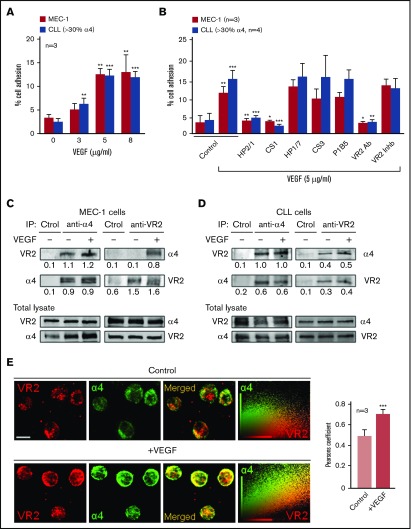
Functional association between α4β1 integrin and VEGFR2 in CLL cells. (A) 2′,7′-Bis-(2-carboxyethyl)-5-(and-6)-carboxyfluorescein acetoxymethyl ester (BCECF-AM)–labeled MEC-1 cells (3 independent experiments) or primary CLL cells from 3 patients (P4, P9, P10) were added to wells coated with the indicated concentrations of VEGF. After 60 minutes at 37°C, attached cells were quantified using a fluorescence analyzer. (B) MEC-1 cells (3 independent experiments) or primary CLL cells (P1, P4, P5, P9), with or without previous incubation with the indicated inhibitors, were added to wells coated with 5 μg/mL VEGF and adhesion was quantified as explained. Values represent the percentage of the total number of cells added. (C-D) A total of 15 × 106 MEC-1 cells (C) or primary CLL cells (D; P4) were serum-starved for 2 hours and treated or not with 50 ng/mL soluble VEGF for 15 minutes. Cells were lysed, immunoprecipitated (IP) with anti-α4, anti-VEGFR2 (VR2), or control (Ctrol) Abs and analyzed by western blotting. The total lysate for each condition was also analyzed by western blotting. Numbers indicate the ratio of immunoprecipitated protein with respect to the amount of that protein in the total lysate. (E) Primary CLL cells (P2, P5, P11) were cultured (2 hours, 37°C) on glass coverslips coated with 10 μg/mL poly-l -lysine (control, top panels) or 5 μg/mL VEGF (bottom panels). Cells were fixed and analyzed by confocal microscopy using the indicated primary Abs and Alexa 568– or Alexa 488–labeled secondary Abs. Colocalization of α4 integrin (green) and VEGFR2 (red) was further demonstrated by dot-plot analyses and quantified by the Pearson correlation coefficient. Scale bar, 4 µm. Confocal images for P11 and average values for the 3 patients analyzed ± standard error of the mean (SEM) are shown. *P < .05; **P ≤ .01; ***P ≤ .001.
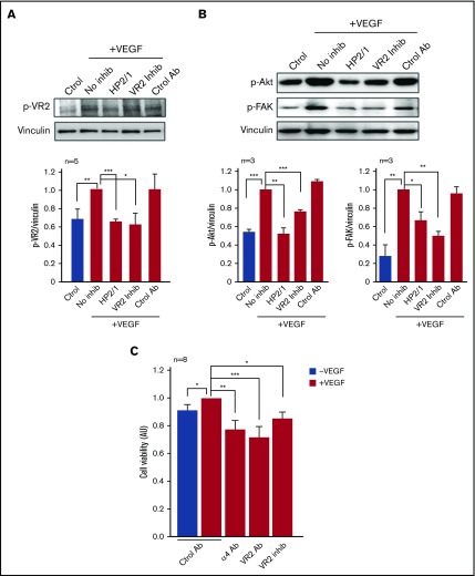
α4β1 integrin contributes to VEGF-induced signaling and function in CLL cells. (A) CLL cells (P13, P14, P15, P16, P17) were serum-starved for 2 hours and treated or not with the indicated inhibitors for 30 minutes at 37°C. Cells were added to wells coated with 5 μg/mL VEGF or 0.1% bovine serum albumin (BSA; control) and after 15 minutes at 37°C, cells were lysed and lysates analyzed by western blotting. VEGFR2 phosphorylation (p-VR2) at Y1214 is shown for a representative sample (P17) and quantitated for all 5 patients (P13, P14, P15, P16, P17) studied. (B) CLL cells (P3, P12, P15) were treated as in panel A and the phosphorylation of Akt and FAK (p-FAK) was analyzed by western blotting. (C) CLL cells (P10, P14, P15, P19, P20, P21, P22, P23) were treated with the indicated Abs for 30 minutes and incubated for 48 hours in the absence (blue bar) or presence (red bars) of immobilized VEGF (5 μg/mL). Cell viability was measured by the CCK8 method. The viability of cells treated with control Ab and VEGF was normalized to 1. Average values (arbitrary units [AU]) ± SEM are shown. *P < .05; **P ≤ .01; ***P ≤ .001.
Similar articles
-
VEGF/VEGFR2 interaction down-regulates matrix metalloproteinase-9 via STAT1 activation and inhibits B chronic lymphocytic leukemia cell migration.Blood. 2010 Jan 28;115(4):846-9. doi: 10.1182/blood-2009-08-239426. Epub 2009 Nov 19. Blood. 2010. PMID: 19965686
-
CLL, but not normal, B cells are dependent on autocrine VEGF and alpha4beta1 integrin for chemokine-induced motility on and through endothelium.Blood. 2005 Jun 15;105(12):4813-9. doi: 10.1182/blood-2004-10-4054. Epub 2005 Feb 24. Blood. 2005. PMID: 15731179
-
A 17-residue sequence from the matrix metalloproteinase-9 (MMP-9) hemopexin domain binds α4β1 integrin and inhibits MMP-9-induced functions in chronic lymphocytic leukemia B cells.J Biol Chem. 2012 Aug 10;287(33):27601-13. doi: 10.1074/jbc.M112.354670. Epub 2012 Jun 22. J Biol Chem. 2012. PMID: 22730324 Free PMC article. Clinical Trial.
-
Functional and Clinical Significance of the Integrin Alpha Chain CD49d Expression in Chronic Lymphocytic Leukemia.Curr Cancer Drug Targets. 2016;16(8):659-668. doi: 10.2174/1568009616666160809102219. Curr Cancer Drug Targets. 2016. PMID: 27514846 Review.
-
Survival of chronic lymphocytic leukemia cells: CD40L and the vascular endothelial growth factor (VEGF) connection.Leukemia. 2005 Apr;19(4):531-2. doi: 10.1038/sj.leu.2403677. Leukemia. 2005. PMID: 15861177 Review. No abstract available.
Cited by
-
Growth Factors VEGF-A165 and FGF-2 as Multifunctional Biomolecules Governing Cell Adhesion and Proliferation.Int J Mol Sci. 2021 Feb 12;22(4):1843. doi: 10.3390/ijms22041843. Int J Mol Sci. 2021. PMID: 33673317 Free PMC article.
-
Comparative Approach to the Temporo-Spatial Organization of the Tumor Microenvironment.Front Oncol. 2019 Nov 7;9:1185. doi: 10.3389/fonc.2019.01185. eCollection 2019. Front Oncol. 2019. PMID: 31788448 Free PMC article. Review.
-
Tumor Cell Survival Factors and Angiogenesis in Chronic Lymphocytic Leukemia: How Hot Is the Link?Cancers (Basel). 2024 Dec 29;17(1):72. doi: 10.3390/cancers17010072. Cancers (Basel). 2024. PMID: 39796700 Free PMC article. Review.
-
Vascular endothelial growth factor signaling in health and disease: from molecular mechanisms to therapeutic perspectives.Signal Transduct Target Ther. 2025 May 19;10(1):170. doi: 10.1038/s41392-025-02249-0. Signal Transduct Target Ther. 2025. PMID: 40383803 Free PMC article. Review.
-
Relation of Neutrophil Gelatinase-Associated Lipocalin Overexpression to the Resistance to Apoptosis of Tumor B Cells in Chronic Lymphocytic Leukemia.Cancers (Basel). 2020 Jul 31;12(8):2124. doi: 10.3390/cancers12082124. Cancers (Basel). 2020. PMID: 32751884 Free PMC article.
References
-
- Lagneaux L, Delforge A, Bron D, De Bruyn C, Stryckmans P. Chronic lymphocytic leukemic B cells but not normal B cells are rescued from apoptosis by contact with normal bone marrow stromal cells. Blood. 1998;91(7):2387-2396. - PubMed
-
- de la Fuente MT, Casanova B, Moyano JV, et al. . Engagement of alpha4beta1 integrin by fibronectin induces in vitro resistance of B chronic lymphocytic leukemia cells to fludarabine. J Leukoc Biol. 2002;71(3):495-502. - PubMed
-
- Lee YK, Bone ND, Strege AK, Shanafelt TD, Jelinek DF, Kay NE. VEGF receptor phosphorylation status and apoptosis is modulated by a green tea component, epigallocatechin-3-gallate (EGCG), in B-cell chronic lymphocytic leukemia. Blood. 2004;104(3):788-794. - PubMed
-
- Till KJ, Spiller DG, Harris RJ, Chen H, Zuzel M, Cawley JC. CLL, but not normal, B cells are dependent on autocrine VEGF and alpha4beta1 integrin for chemokine-induced motility on and through endothelium. Blood. 2005;105(12):4813-4819. - PubMed
-
- Shanafelt TD, Kay NE. The clinical and biologic importance of neovascularization and angiogenic signaling pathways in chronic lymphocytic leukemia. Semin Oncol. 2006;33(2):174-185. - PubMed
