. 2019 Sep 1;79(17):4426-4438.
doi: 10.1158/0008-5472.CAN-19-0237. Epub 2019 Jul 15.

APE1 Upregulates MMP-14 via Redox-Sensitive ARF6-Mediated Recycling to Promote Cell Invasion of Esophageal Adenocarcinoma

Affiliations

APE1 Upregulates MMP-14 via Redox-Sensitive ARF6-Mediated Recycling to Promote Cell Invasion of Esophageal Adenocarcinoma

Heng Lu et al. Cancer Res. .

Abstract

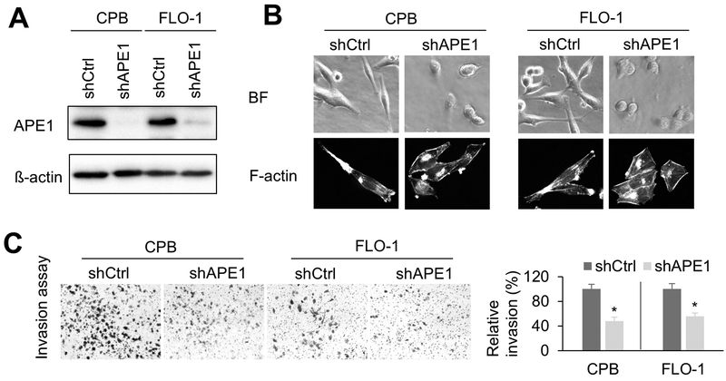
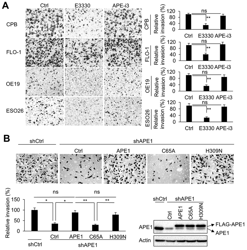
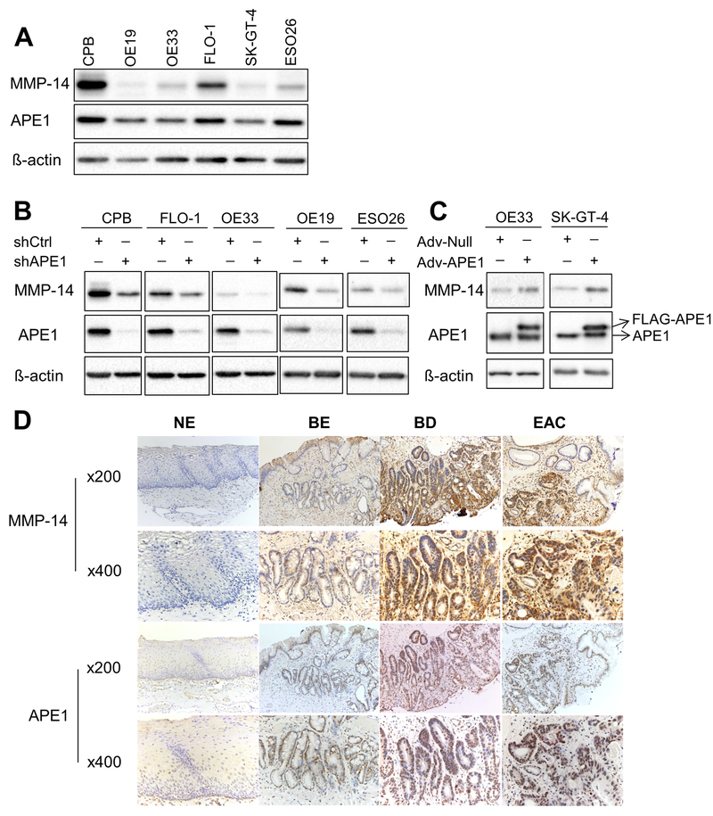
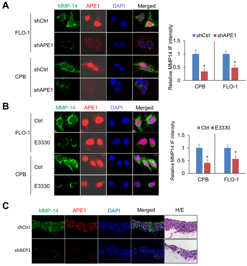
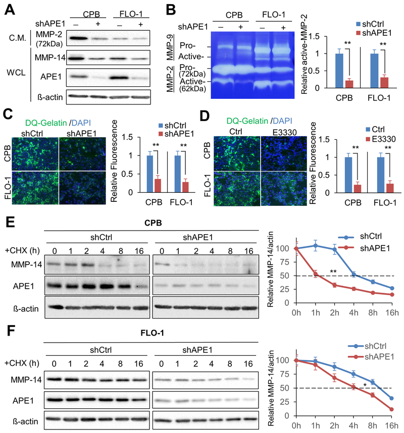
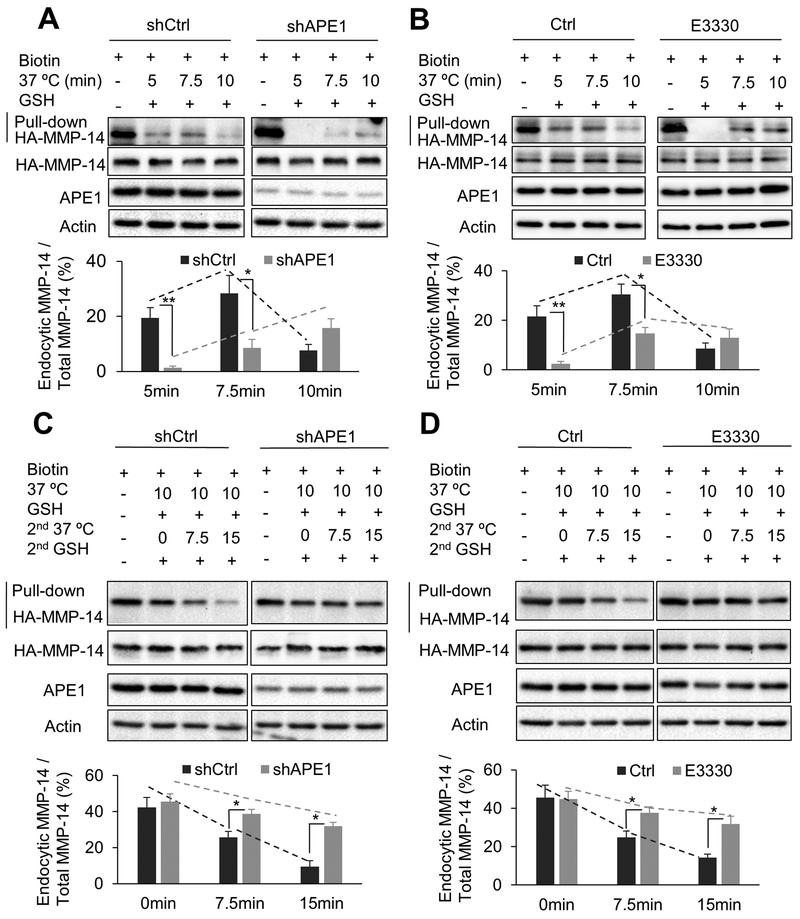
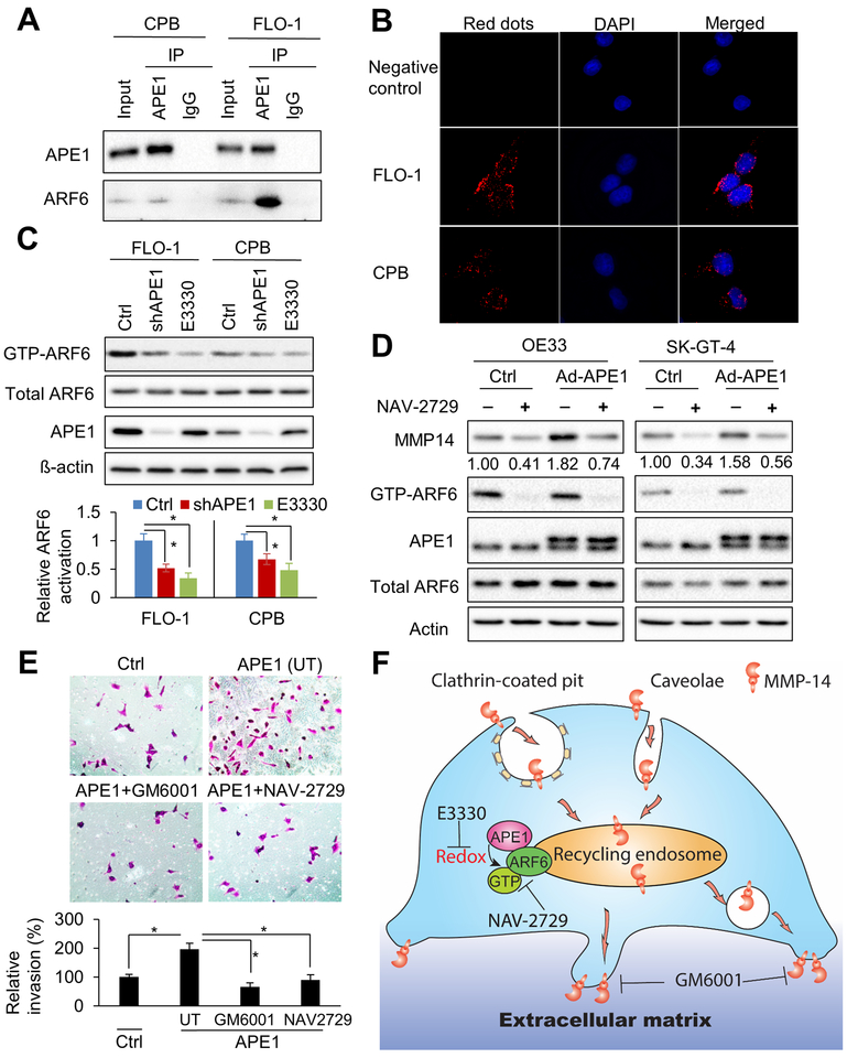
Esophageal adenocarcinoma (EAC) is an aggressive malignancy with poor clinical outcome. The incidence of EAC has been rising rapidly in the past three decades. Here, we showed that apurinic/apyrimidinic endonuclease (APE1) is overexpressed in EAC cell lines, and patients' samples of dysplasia and EAC. Downregulation of APE1 or inhibition of its redox function significantly repressed invasion. Overexpression of a redox-defective mutant, C65A, abrogated the proinvasive phenotype of APE1. APE1 regulated invasion via upregulation of matrix metalloproteinase 14 (MMP-14), which subsequently activated MMP-2, leading to degradation of the extracellular matrix in a redox-dependent manner. Downregulation of APE1 or inhibition of its redox function decreased the rate of endocytosis and recycling of MMP-14 protein. APE1 interacted with ARF6, a key regulator of MMP-14 recycling, which maintained ARF6 activity in an APE1-redox-dependent manner, promoting its ability to regulate MMP-14 recycling to the cell surface. In summary, these findings identify a novel redox-sensitive APE1-ARF6-MMP-14 signaling axis that mediates cellular invasion in esophageal carcinogenesis. SIGNIFICANCE: This study demonstrates the association between oxidative stress and the development and metastatic behavior of esophageal adenocarcinoma.


Conflict of interest statement

Disclosure of Potential Conflicts of Interest:

No potential conflicts of interest were disclosed.

Figures

Figure1
Figure1. silencing decreases invasion capacity.
A, Western blot analysis of CPB and FLO-1 cells with APE1-knockdown (shAPE1) or control (shCtrl). B, Representative cell images of bright field (BF) and F-actin staining in APE1-knockdown cells or control cells. Alexa Fluor™ 488 Phalloidin was used for F-actin staining. C, invasion assays were performed using APE1-knockdown cells or respective control cells. The results were expressed as the mean ± SEM of three independent experiments. *, p < 0.05.
Figure 2.
Figure 2.. Redox function of APE1 is required for invasion.
A, Representative images (magnification: 200x) of the invasion assays. CPB, FLO-1, OE19, and ESO26 cells were pre-treated overnight with 40μM E3330 (APE1 redox function inhibitor), 0.5μM APE1-iIII (APE1 DNA repair function inhibitor), or vehicle control (Ctrl), followed by collecting cells for invasion assays. The inhibitors were maintained in the invasion chambers and bottom wells during the assays. B, Representative images (magnification: 200x) of the invasion assays in FLO-1 cells. Control vector (Ctrl), FLAG-tagged wild-type APE1, C65A (redox defective mutant) or H309N (DNA repair defective mutant) was overexpressed in APE1-knockdown (shAPE1) FLO-1 cells. The mutation to avoid APE1-shRNA targeting has been generated in those overexpression vectors. Scramble shRNA was used as control (shCtrl). Endogenous APE1, overexpression of FLAG-tagged APE1 and its relevant mutants were examined by Western blot analysis. All quantification analyses were based on independent triplicate experiments. *, p < 0.05; **, p < 0.01; ns, no significance.
Figure 3.
Figure 3.. MMP-14 protein level is regulated by APE1 in BE and EAC cell lines.
A, Western blot analysis of APE1 and MMP-14 protein profiles in dysplastic Barrett’s cell line (CPB) and EAC cell lines. B, MMP-14, APE1 and ß-actin protein levels were examined by Western blot analysis in CPB, FLO-1, OE33, OE19 and ESO26 cells with stable APE1 knockdown (shAPE1). Stable scramble shRNA (shCtrl) was used as the control. C, APE1 was overexpressed in OE33 and SK-GT-4 cells by using adenoviral infection particles (Adv-APE1). Adv-Null was used as the control. The protein samples were collected 72 h post infection for Western blot analysis of MMP-14, APE1, and ß-actin. D, Representative immunohistochemistry staining images of MMP-14 and APE1 in normal esophagus (NE), non-dysplastic Barrett’s esophagus (BE), dysplastic Barrett’s esophagus (BD) and esophageal adenocarcinoma (EAC).
Figure 4.
Figure 4.. APE1 silencing or APE1 redox-specific inhibition decreases MMP-14 protein levels.
Representative immunofluorescent images of MMP-14 (green) and APE1 (red) in FLO-1 or CPB cells; nuclei were stained with DAPI (blue). A, APE1 stable knockdown cells (shAPE1), and their control cells (shCtrl). B, Cells were treated with 40μM E3330 or vehicle control (Ctrl) for 24 h before staining. C, 3D organotypic culture of CPB cells with stable knockdown (shAPE1) or control (shCtrl). Hematoxylin and eosin (H&E) staining of sequential cut of the same blocks used for immunofluorescence staining. The quantification of MMP-14 intensity was expressed as the mean ± SEM of 3 independent fields. *, p < 0.05.
Figure 5.
Figure 5.. Knockdown of APE1 reduces active MMP-2 form and ECM degradation by down-regulating MMP-14 protein level.
A, Conditioned medium (C.M.) from CPB, FLO-1 or relevant APE1 knockdown cells were collected, concentrated, and examined by Western blot analysis for MMP-2. The whole cell lysates from the relevant dishes were collected for Western blots to detect MMP-14, APE1 and ß-actin. B, In-gel zymography assay. The concentrated conditioned medium was loaded in gelatin gel to test MMP-2 activities. Arrows indicate pro- or active- forms of MMPs. Active MMP-2 levels were measured by the intensity ratio of active MMP-2 / actin. C, In situ zymography assay was performed by using APE1 knockdown cells or control cells on DQ-gelatin pre-coated slides at 48 h post cell seeding. Relative active MMP-2 activities were measured by the active MMP-2/ß-actin. D, In situ zymography assay by the cells treated with 40μM E3330 or vehicle control (Ctrl). ECM degradation activities were measured by immunofluorescent intensities. E & F, Cycloheximide (CHX) chase assays were performed by using 100μg/ml cycloheximide in CPB (E) and FLO-1 (F) cells. The cell lysates were collected at the indicated time points for Western blot analysis of APE1 knockdown cells (shAPE1) or control cells (shCtrl). Measurement of MMP-14 protein stability was based on the intensity ratio of MMP-14/actin. All quantification analyses were based on independent triplicate experiments. *, p < 0.05; **, p < 0.01.
Figure 6.
Figure 6.. APE1 is required for the rapid MMP-14 endocytic recycling in EAC cells.
A & B, Endocytosis assays of HA-tagged MMP-14 in FLO-1 cells. Cells were transfected with HA-tagged MMP-14. The cell lysates were collected for biotin pulldown at the indicated time points of endocytosis (37°C incubation). C & D, Recycling assays of HA-tag labelled MMP-14 in FLO-1 cells. Recycling assays were performed after 10 min endocytosis (37°C incubation). The cell lysates were collected for biotin pulldown at indicated time courses of recycling (2nd 37°C incubation). APE1-knockdown cells (shAPE1) and control cells (shCtrl) of FLO-1 were used (A, C). FLO-1 cells were pretreated with 40μM E3330 for 24 h before experiments (B, D). Relative endocytic MMP-14 levels were measured by the ratio of endocytic MMP-14/total MMP-14. All quantification analyses were based on independent triplicate experiments. *, p < 0.05; **, p < 0.01.
Figure 7.
Figure 7.. APE1 promotes ARF6 activity through interaction and redox function.
A, immunoprecipitation (IP) of APE1 in CPB and FLO-1 cells. IgG immunoprecipitation works as a negative control. Western blots of APE1 and ARF6 were performed. B, Proximity ligation assays (PLA) in FLO-1 and CPB cells were performed by using anti-ARF6 and anti-APE1 antibodies. Single antibody (anti-ARF6) was used as negative control. C, ARF6 activity assays in CPB and FLO-1 cells. The active ARF6 form, GTP-ARF6, was specifically pulled down from whole cell lysates by GGA3 PBD agarose beads. APE1, total ARF6, and ß-actin in whole cell lysates, and GTP-ARF6 in pulldown products were examined by Western blots. Stable APE1-knockdown cells (shAPE1), control cells (Ctrl) or the cells with 24 h pretreatment of 40μM E3330 were harvested for cell lysates. Relative ARF6 activation was measured by the ratio of (GTP-ARF6)/total ARF6. D, APE1 was overexpressed in OE33 and SK-GT-4 cells by adenoviral infection (Ad-APE1). Cells were treated with 1μM specific ARF6 inhibitor, NAV-2729, for 48 h. Whole cell lysates were collected for GTP-ARF6 pulldown and Western blot analysis. E, Representative images (magnification: 200x) of the invasion assays in SK-GT-4 cells. Control vector (Ctrl), or wild-type APE1 was overexpressed in the cells. APE1-overexpressed cells were pre-treated and maintained with 10μM MMPs inhibitor, GM6001, or 1uM NAV-2729 through invasion assay. UT, untreated. All quantification analyses were based on independent triplicate experiments. *, p < 0.05. F, schematic summary of APE1-mediated MMP-14 endocytosis and recycling. MMP-14 is efficiently internalized from plasma membrane by clathrin-dependent or caveolar endocytosis. Active endocytosis and recycling of MMP-14 is critical for extracellular matrix (ECM) degradation and cell invasion. APE1 promotes ARF6 activation through interaction and APE1-redox function. Active ARF6 (GTP-ARF6) in recycling endosome drives quick recycling of MMP-14. APE1 redox inhibitor, E3330, or ARF6 specific inhibitor, NAV-2729, can significantly decrease MMP-14 recycling to cell surface and subsequent cell invasion. Moreover, MMPs inhibitor, GM6001 can inhibit MMP-14 and MMP-2 activities and repress cell invasion.

References

    1. Jemal A, Siegel R, Xu J, Ward E. Cancer statistics, 2010. CA Cancer J Clin 2010;60:277–300 - PubMed
    1. Kong CY, Kroep S, Curtius K, Hazelton WD, Jeon J, Meza R, et al. Exploring the recent trend in esophageal adenocarcinoma incidence and mortality using comparative simulation modeling. Cancer Epidemiol Biomarkers Prev 2014;23:997–1006 - PMC - PubMed
    1. Sital RR, Kusters JG, De Rooij FW, Kuipers EJ, Siersema PD. Bile acids and Barrett’s oesophagus: a sine qua non or coincidence? Scand J Gastroenterol Suppl 2006:11–7 - PubMed
    1. Bernstein H, Bernstein C, Payne CM, Dvorak K. Bile acids as endogenous etiologic agents in gastrointestinal cancer. World J Gastroenterol 2009;15:3329–40 - PMC - PubMed
    1. Lagergren J Adenocarcinoma of oesophagus: what exactly is the size of the problem and who is at risk? Gut 2005;54 Suppl 1:i1–5 - PMC - PubMed

Publication types

MeSH terms

Substances

Supplementary concepts