. 2019 Oct;30(10):1886-1897.
doi: 10.1681/ASN.2019020192. Epub 2019 Jul 15.

Glomerular Function and Structural Integrity Depend on Hyaluronan Synthesis by Glomerular Endothelium

Affiliations

Glomerular Function and Structural Integrity Depend on Hyaluronan Synthesis by Glomerular Endothelium

Bernard M van den Berg et al. J Am Soc Nephrol. 2019 Oct.

Abstract

Background: A glycocalyx envelope consisting of proteoglycans and adhering proteins covers endothelial cells, both the luminal and abluminal surface. We previously demonstrated that short-term loss of integrity of the luminal glycocalyx layer resulted in perturbed glomerular filtration barrier function.

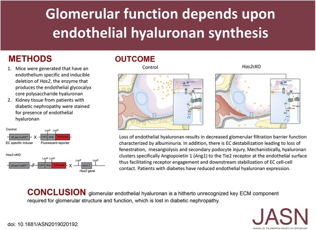
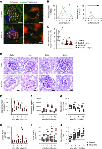
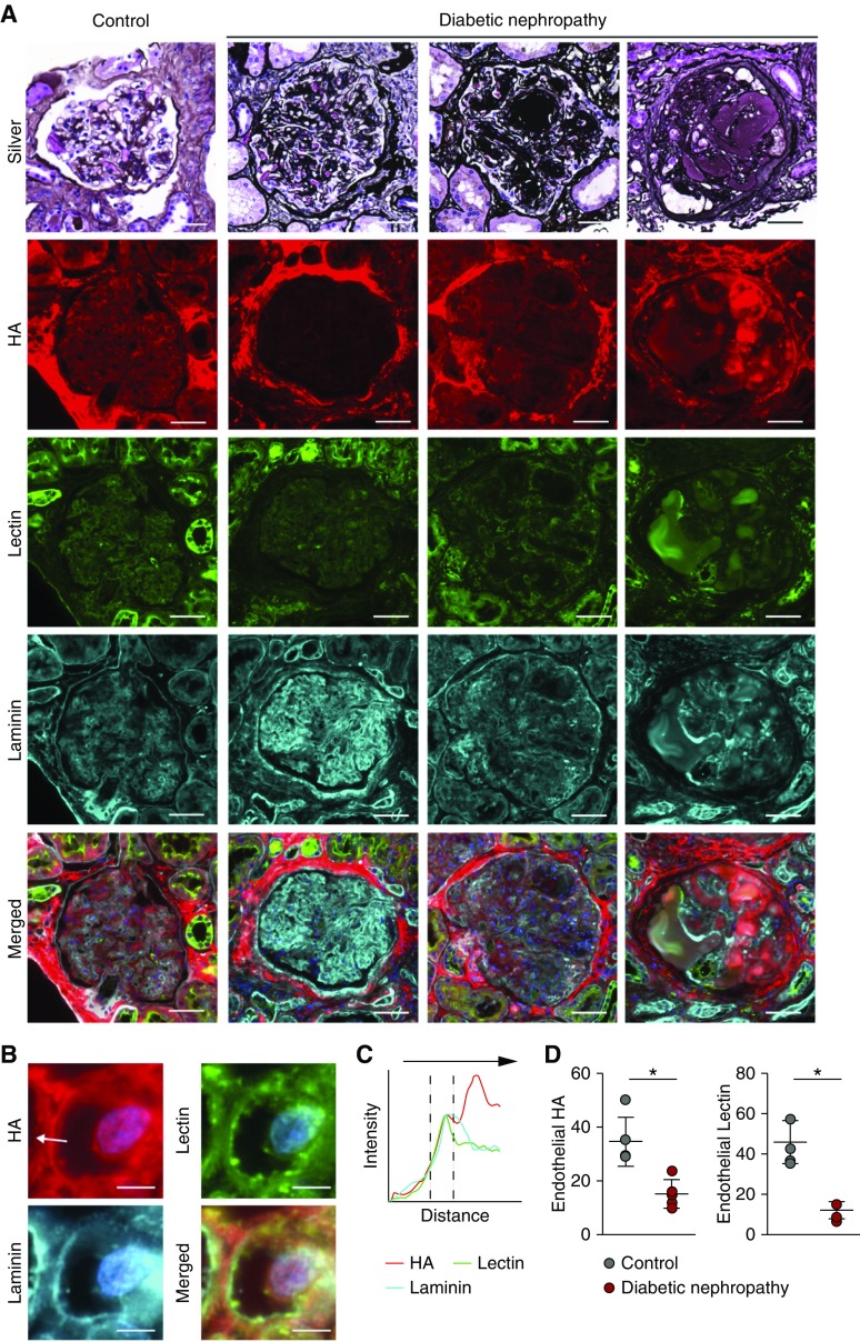
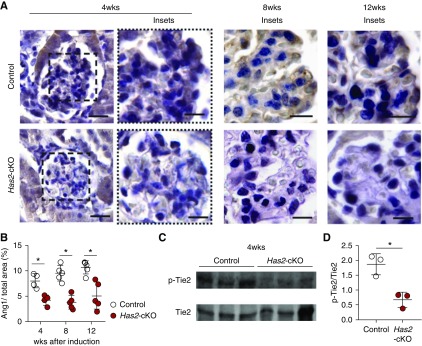
Methods: To explore the role of the glycocalyx layer of the endothelial extracellular matrix in renal function, we generated mice with an endothelium-specific and inducible deletion of hyaluronan synthase 2 (Has2), the enzyme that produces hyaluronan, the main structural component of the endothelial glycocalyx layer. We also investigated the presence of endothelial hyaluronan in human kidney tissue from patients with varying degrees of diabetic nephropathy.

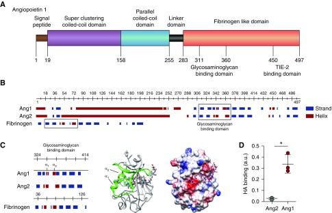
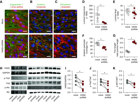
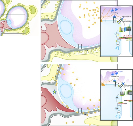
Results: Endothelial deletion of Has2 in adult mice led to substantial loss of the glycocalyx structure, and analysis of their kidneys and kidney function showed vascular destabilization, characterized by mesangiolysis, capillary ballooning, and albuminuria. This process develops over time into glomerular capillary rarefaction and glomerulosclerosis, recapitulating the phenotype of progressive human diabetic nephropathy. Using a hyaluronan-specific probe, we found loss of glomerular endothelial hyaluronan in association with lesion formation in tissue from patients with diabetic nephropathy. We also demonstrated that loss of hyaluronan, which harbors a specific binding site for angiopoietin and a key regulator of endothelial quiescence and maintenance of EC barrier function results in disturbed angiopoietin 1 Tie2.

Conclusions: Endothelial loss of hyaluronan results in disturbed glomerular endothelial stabilization. Glomerular endothelial hyaluronan is a previously unrecognized key component of the extracelluar matrix that is required for glomerular structure and function and lost in diabetic nephropathy.

Keywords: angiopoietin-1; glomerular endothelial cells; glomerulosclerosis; glycocalyx; hyaluronan.


Figures

None
Graphical abstract
Figure 1.
Figure 1.
Loss of endothelial HA and glycocalyx results in progressive glomerulopathy and proteinuria. (A) Left, representative cross-sectional confocal images of glomerular tissue sections, 4 weeks after tamoxifen induction. Sections were stained with the HA-binding probe Ncan-eGFP (green), and successfully recombined ECs were labeled with TdTomato (red). Has2-cKO mice show loss of the endothelial HA layer (scale bar, 30 µm). Right, examples of measurement lines over glomerular free capillary wall (scale bar, 5 µm). (B) Fluorescence intensity plots demonstrate intraluminal (green, left) and subendothelial (green, right) localization of HA in relation to the EC membrane position (red, dotted lines). (C) Luminal HA surface coverage quantification (n=3 per group). (D) Representative images of periodic acid–Schiff–stained glomeruli of control (upper panels) and Has2-cKO (lower panels) mice at 2, 4, 8, and 12 weeks after tamoxifen induction. Arrowheads indicate diffuse widening of capillary loops through mesangiolysis (ballooning; scale bar, 50 µm). (E) Glomerular capillary surface area increased from week 2 onwards. (F) At this early stage, glomerular hypertrophy was the most prominent change in Has2-cKO glomeruli, which was accompanied by induction of matrix expansion. (G) From week 4 onwards capillary rarefaction developed; (E–G) 2 (n=4, 5 per group), 4 (n=6, 4 per group), 8 (n=5 group), and 12 weeks (n=5 group). (H) Albumin-to-creatinine ratio (ACR) in 24-hour urine samples significantly increased in Has2-cKO from week 4. During the 12-week period after tamoxifen induction, (I) Has2-cKO mice drank more water (no change in food intake) and (J) total body weight significantly increased from week 8 onward; 2 (n=8, 12 per group), 4 (n=14 per group), 8 (n=10 per group), and 12 weeks (n=5 per group). Values are given as (C) mean±SEM or (EJ) mean±SD. Difference was assessed by nonpaired two-tailed t test or, when not normally distributed, by two-tailed F-test; *P<0.05. FL, fluorescence intensity.
Figure 2.
Figure 2.
Early-stage human diabetic nephropathy shows loss of glomerular HA. (A) Representative methenamine silver–periodic acid–Schiff–stained glomeruli of a control glomerulus (left panel) and three consecutive stages of DN in human kidney biopsy specimens. Further, fluorescent images of glomerular tissue sections stained for HA (red, second row), Lycopersicon esculentum lectin (green, third row), and laminin (cyan, fourth row), and merged images. (B) Insets show examples of measurement lines (scale bar, 5 µm). (C) Fluorescence intensity plots demonstrate endothelial localization of HA in relation to the subendothelial basement membrane position (cyan, laminin). (D) Quantification of total endothelial HA (left) or lectin (right) presence (n=5 per group).
Figure 3.
Figure 3.
HA binds angiopoietin 1. (A) Structural characteristics of angiopoietin 1 (Ang1) reveal, besides the superclustering coiled-coil domain (purple) and parallel coiled-coil domain (blue) allowing Ang1 molecules to form clusters, a possible glycosaminoglycan- (GAG) binding domain within the N-terminal fibrinogen-like domain (orange) that neighbors the TIE2-binding site. (B) In contrast to the predicted GAG-binding domain (right box) of angiopoietin 2 (Ang2), the Ang1 GAG-binding domain resembles the HA-binding domain of fibrinogen (left box). (C) Like the predicted HA-binding Link module superfamily, the Ang1 GAG-binding domain contains two α-helices separated by an antiparallel β strand (left panel). The 3D ribbon configuration of the fibrinogen-like domain of Ang1 shows the position of the two α-helices in relation to the proposed HA-binding domain (middle panel), and the electrostatic surface projection of the 3D crystallographic structure reveals the positions of negative- (red), neutral- (white), and positive- (blue) charged sites within the HA-binding domain (right panel). (D) Direct binding of recombinant human Ang1 or Ang2 to HA. Values are given as mean±SD and difference was assessed by nonpaired two-tailed t test; *P<0.05.
Figure 4.
Figure 4.
HA angiopoietin 1 clustering stabilizes the endothelial layer. Representative cross-sectional confocal images of primary human glomerular-derived microvascular ECs (hgMVECs), transduced with an empty- (mock) or a HAS2-shRNA–containing lenti-viral construct (HAS2 shRNA) and exposed to a laminar shear stress of 5 dyne/cm2 for 3 days in the presence of 100 ng/ml recombinant human Ang1, stained (A) for HA (red) and Ang1 (green), revealed that especially at cell-cell contact areas (arrow) Ang1 presence is reduced, which is accompanied with (B) reduced membrane-bound active β-catenin (anti-ABC, green) and (C) increased unstable focal adherence junctions (FAJ) as visualized by VE-cadherin (CD144, green) staining. Both in combination with fluorescent-labeled phallotoxin (phalloidin-TRITC, red) to visualize intracellular F-actin and Hoechst 33258 (blue) nuclear stain (scale bar, 20 µm). Silencing of HAS2 results in reduced (D) HA expression, (E) Ang1 binding, (F) membrane-bound β-catenin, and (G) induction of unstable focal adherence junctions (FAJ) (n=5 per group). (H) Western-blot samples stained for HAS2, GAPDH, p-Tie2, Tie2, p-Akt, and Akt. Quantification of (I) HAS2 gene expression, (J) p-Tie2(Y992) over Tie2 expression, and (K) the downstream signaling Akt activation. Values are given as mean±SEM (D–G) or as individual data points (I–K). Differences between control and HAS2-shRNA–treated cells were assessed by paired two-tailed t test; *P<0.05.
Figure 5.
Figure 5.
Loss of HA leads to reduction of Ang1/Tie2 signaling in vivo. (A) Representative images of glomerular angiopoietin 1 (Ang1) presence at 4 weeks (left panels) after tamoxifen induction in control (upper panels) and has2-cKO (lower panels) mice (scale bar, 60 µm) with detailed glomerular tufts at 4, 8, and 12 weeks (scale bar, 30 µm). (B) Quantification of glomerular tuft Ang1 (n=5 per group). (C) Western blot samples from kidneys of Has2-cKO and control mice at 4 weeks stained for p-Tie2 and Tie2. (D) Quantification of p-Tie2(Y992) over Tie2 expression. Values are given as mean±SEM or mean±SD and difference was assessed by nonpaired two-tailed t test; *P<0.05.
Figure 6.
Figure 6.
Glomerular integrity depends on endothelial surface HA. Schematic overview of loss of endothelial HA results in decreased glomerular filtration barrier function characterized by albuminuria (yellow dots). In addition, there is EC destabilization leading to loss of fenestration, mesangiolysis (yellow asterisk), and secondary podocyte injury. Mechanistically, HA clusters specifically angiopoietin 1 (Ang1) to the TIE2 receptor at the endothelial surface, thus facilitating receptor engagement and downstream stabilization of EC cell-cell contact.

References

    1. Dane MJ, van den Berg BM, Lee DH, Boels MG, Tiemeier GL, Avramut MC, et al. : A microscopic view on the renal endothelial glycocalyx. Am J Physiol Renal Physiol 308: F956–F966, 2015 - PubMed
    1. Sarrazin S, Lamanna WC, Esko JD: Heparan sulfate proteoglycans. Cold Spring Harb Perspect Biol 3: 1–33, 2011 - PMC - PubMed
    1. Bishop JR, Stanford KI, Esko JD: Heparan sulfate proteoglycans and triglyceride-rich lipoprotein metabolism. Curr Opin Lipidol 19: 307–313, 2008 - PubMed
    1. Moretto P, Karousou E, Viola M, Caon I, D’Angelo ML, De Luca G, et al.: Regulation of hyaluronan synthesis in vascular diseases and diabetes. J Diabetes Res 2015: 1–9, 2015 - PMC - PubMed
    1. Viola M, Karousou E, D’Angelo ML, Caon I, De Luca G, Passi A, et al.: Regulated hyaluronan synthesis by vascular cells. Int J Cell Biol 2015: 1–8, 2015 - PMC - PubMed

Publication types

Substances

LinkOut - more resources