. 2019 Oct;18(10):1744-1754.
doi: 10.1158/1535-7163.MCT-19-0156. Epub 2019 Jul 15.

Targeting MUC1-C Inhibits TWIST1 Signaling in Triple-Negative Breast Cancer

Affiliations

Targeting MUC1-C Inhibits TWIST1 Signaling in Triple-Negative Breast Cancer

Tsuyoshi Hata et al. Mol Cancer Ther. 2019 Oct.

Abstract

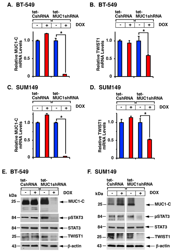
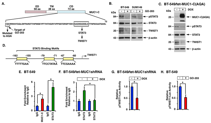
The oncogenic MUC1-C protein and the TWIST1 epithelial-mesenchymal transition transcription factor (EMT-TF) are aberrantly expressed in triple-negative breast cancer (TNBC) cells. However, there is no known association between MUC1-C and TWIST1 in TNBC or other cancer cells. Here, we show that MUC1-C activates STAT3, and that MUC1-C and pSTAT3 drive induction of the TWIST1 gene. In turn, MUC1-C binds directly to TWIST1, and MUC1-C/TWIST1 complexes activate MUC1-C expression in an autoinductive circuit. The functional significance of the MUC1-C/TWIST1 circuit is supported by the demonstration that this pathway is sufficient for driving (i) the EMT-TFs, ZEB1 and SNAIL, (ii) multiple genes in the EMT program as determined by RNA-seq, and (iii) the capacity for cell invasion. We also demonstrate that the MUC1-C/TWIST1 circuit drives (i) expression of the stem cell markers SOX2, BMI1, ALDH1, and CD44, (ii) self-renewal capacity, and (iii) tumorigenicity. In concert with these results, we show that MUC1-C and TWIST1 also drive EMT and stemness in association with acquired paclitaxel (PTX) resistance. Of potential therapeutic importance, targeting MUC1-C and thereby TWIST1 reverses the PTX refractory phenotype as evidenced by synergistic activity with PTX against drug-resistant cells. These findings uncover a master role for MUC1-C in driving the induction of TWIST1, EMT, stemness, and drug resistance, and support MUC1-C as a highly attractive target for inhibiting TNBC plasticity and progression.


Conflict of interest statement

Potential Conflict of Interest: DK has equity interests in Genus Oncology, Reata Pharmaceuticals, Hillstream BioPharma, Nanogen Therapeutics and Victa BioTherapeutics, serves as a member of the board of directors of Genus, Nanogen and Victa, and is a paid consultant to Genus, Reata, CanBas and Victa. The other authors disclosed no potential conflicts of interest.

Figures

Figure 1.
Figure 1.. Targeting MUC1-C suppresses TWIST1 expression.
A-F. BT-549 and SUM149 cells stably expressing a tet-CshRNA or tet-MUC1shRNA were left untreated or treated with 500 ng/ml DOX for 7 days. Cells were analyzed for MUC1-C (A,C) and TWIST1 (B,D) mRNA levels by qRT-PCR using primers listed in Supplemental Table S1. The results (mean±SD) are expressed as relative mRNA levels compared to that obtained for control untreated cells (assigned a value of 1). The asterisk (*) denotes a p-value of <0.05. Lysates were immunoblotted with antibodies against the indicated proteins (E,F).
Figure 2.
Figure 2.. MUC1-C drives TWIST1 activation.
A. Schema of MUC1-C with the extracellular domain (ED), transmembrane (TM) region and amino acid sequence of the intrinsically disordered cytoplasmic domain (CD). Highlighted is the CQC motif, which is necessary for MUC1-C homodimerization and nuclear import. Mutation of the CQC motif to AQA inactivates MUC1-C function. The CQC motif is the target of the cell-penetrating GO-203 inhibitor (R9-CQCRRKN). Also highlighted is the STAT3 binding site. B. BT-549 (left) and SUM149 (right) cells were left untreated or treated with 5 μM GO-203 for 48 h. Lysates were immunoblotted with antibodies against the indicated proteins. C. BT-549 cells expressing a tet-MUC1-C(AQA) vector were left untreated or treated with 500 ng/ml DOX for 7 days. Lysates were analyzed by immunoblotting with antibodies against the indicated proteins. D. Schema of the TWIST1 promoter with depiction of putative STAT3 binding motifs. E. Soluble chromatin from BT-549 cells was precipitated with anti-MUC1-C, anti-STAT3 or a control IgG. The DNA samples were amplified by qPCR with primers for the TWIST1 promoter (Supplemental Table S2). The results (mean±SD of three determinations) are expressed as the relative fold enrichment compared to that obtained with the IgG control (assigned a value of 1). F. BT-549/tet-MUC1shRNA cells were left untreated or treated with 500 ng/ml DOX for 7 days. Soluble chromatin was precipitated with anti-STAT3 or a control IgG. The DNA samples were amplified by qPCR with primers for the TWIST1 promoter. The results (mean±SD of three determinations) are expressed as the relative fold enrichment compared to that obtained with the IgG control (assigned a value of 1). G and H. BT-549/tet-MUC1shRNA cells were left untreated or treated with 500 ng/ml DOX for 5 days (G). BT-549 cells were left untreated or treated with 5 μM GO-203 for 48 h (H). The cells were transfected with a pTWIST1-Luc reporter for 48 h and then analyzed for Luc activity. The results (mean±SD) are expressed as relative Luc activity compared to that obtained for control untreated cells (assigned a value of 1).
Figure 3.
Figure 3.. MUC1-C forms a complex with TWIST1 on the MUC1 promoter.
A. Nuclear lysates were incubated with anti-MUC1-C or a control IgG. The input and precipitates were analyzed by immunoblotting with antibodies against the indicated proteins. B and C. Purified recombinant TWIST1 was incubated with GST or the indicated GST-MUC1-CD fusion proteins. Precipitates obtained with glutathione beads were immunoblotted with anti-TWIST1. Loading of the GST proteins was analyzed by Coomassie Blue staining. D. Schema of the MUC1 promoter with highlighting of the putative TWIST1 binding motifs. E. Soluble chromatin was precipitated with anti-MUC1-C, anti-TWIST1 or a control IgG (left). Soluble chromatin was precipitated with anti-MUC1-C (ChIP) and then re-precipitated with anti-TWIST1 or a control IgG (re-ChIP)(right). The DNA samples were amplified by qPCR with primers for the MUC1 promoter. The results (mean±SD of three determinations) are expressed as the relative fold enrichment compared to that obtained with the IgG control (assigned a value of 1). F. BT-549/tet-MUC1shRNA cells were left untreated or treated with 500 ng/ml DOX for 7 days. Soluble chromatin was precipitated with anti-TWIST1 or a control IgG. The DNA samples were amplified by qPCR with primers for the MUC1 promoter. The results (mean±SD of three determinations) are expressed as the relative fold enrichment compared to that obtained with the IgG control (assigned a value of 1). G. BT-549/CshRNA and BT-549/TWIST1shRNA cells were analyzed for TWIST1 and MUC1-C mRNA levels by qRT-PCR. The results (mean±SD) are expressed as relative mRNA levels compared to that obtained for control cells (assigned a value of 1).
Figure 4.
Figure 4.. MUC1-C promotes EMT and stemness.
A and B. BT-549/tet-MUC1shRNA cells left untreated or treated with 500 ng/ml DOX for 7 days were analyzed for ZEB1 and SNAIL mRNA levels. The results (mean±SD) are expressed as relative mRNA levels compared to that obtained for control cells (assigned a value of 1)(left). Lysates were analyzed by immunoblotting (right). B. RNA-seq was performed in triplicate on (i) BT-549/CshRNA and BT-549/MUC1shRNA, and (ii) SUM149/CshRNA and SUM149/MUC1shRNA cells. The datasets were analyzed using the Hallmark gene signature collection. EMT was identified as the pathway with the highest association (Supplemental Figs. S5 and S6). C. Lysates from BT-549/tet-MUC1shRNA cells left untreated or treated with 500 ng/ml DOX were immunoblotted with antibodies against the indicated proteins. D. Lysates from BT-549/CshRNA and BT-549/TWIST1shRNA cells were immunoblotted with antibodies against the indicated proteins. E. BT-549/tet-MUC1shRNA cells left untreated (blue bars) or treated with 500 ng/ml DOX (red bars) for 7 days were analyzed for colony formation. The results (mean±SD of at least three determinations) are expressed as the number of colonies/field. F. BT-549/tet-MUC1shRNA cells left untreated (blue bars) or treated with 500 ng/ml DOX (red bars) for 7 days were analyzed for mammosphere formation. The results (mean±SD of at least three determinations) are expressed as the number of mammospheres/well. G. Six-week old nude female mice were injected subcutaneously in the flank with 3 × 10⁶ SUM149/tet-MUC1shRNA cells. Mice were pair-matched into two groups when tumor volumes reached 100–150 mm3 and were fed without (blue circles) and with DOX (red circles). Tumor volumes are expressed as the mean±SD for 6 mice. H. Lysates from tumors harvested on day 21 were immunoblotted with antibodies against the indicated proteins.
Figure 5.
Figure 5.. MUC1-C drives drug resistance.
A. Lysates from drug naïve (WT) BT-549 and BT-549/PTX-R cells were immunoblotted with antibodies against the indicated proteins. B. BT-549 WT and PTX-R cells were analyzed for nuclear MUC1-C and TWIST1 localization by IF microscopy. C and D. Chromatin from BT-549 WT and PTX-R cells was precipitated with anti-MUC1-C, anti-TWIST1 or a control IgG. The DNA samples were amplified by qPCR with primers for the MUC1 (C) and ABCB1 (D) promoters. The results (mean±SD of three determinations) are expressed as the relative fold enrichment compared to that obtained with the IgG control (assigned a value of 1). E. Lysates from BT-549/PTX-R/tet-MUC1shRNA cells left untreated or treated with 500 ng/ml DOX for 7 days were analyzed by immunoblotting. F. BT-549/PTX-R/tet-MUC1shRNA cells were left untreated or treated with 500 ng/ml DOX for 7 days. The cells were then exposed to vehicle control (CTR) or 30 μM PTX for 72 h. The results (mean±SD) are expressed as relative survival as compared to that obtained with DOX-untreated/CTR cells (assigned a value of 1).
Figure 6.
Figure 6.. Targeting MUC1-C reverses PTX resistance and decreases self-renewal capacity.
A. Lysates from BT-549/PTX-R cells left untreated or treated with 5 μM GO-203 for 48 h were analyzed by immunoblotting. B and C. BT-549/PTX-R cells were left untreated or treated with 0.1, 0.3 or 1.0 μM GO-203 in the absence or presence of the indicated PTX concentrations. Cell viability (mean±SD of at least three determinations) was determined by Alamar blue staining (B) and was used to calculate the CI values (C), which at <1.0 denote synergism. D-F. BT-549 drug-naïve (WT) and BT-549/PTX-R cells (D), BT-549/PTX-R/tet-MUC1shRNA cells left untreated or treated with 500 ng/ml DOX for 7 days (E) and BT-549/PTX-R cells left untreated and treated with 5 μM GO-203 for 7 days (F) were analyzed for mammosphere formation. The results (mean±SD of at least three determinations) are expressed as the number of mammospheres/well. G. Proposed model for involvement of MUC1-C in integrating TWIST1, EMT, stemness and drug resistance. MUC1-C activates the STAT3 pathway and is necessary for TWIST1 activation. In turn, MUC1-C binds directly to TWIST1 and forms an autoregulatory MUC1-C→TWIST1 circuit. Importantly, our results show that this circuit is sufficient for driving (i) ZEB1 and SNAIL expression, (ii) multiple genes associated with the EMT program and invasion, (iii) BC stem cell markers, (iv) self-renewal and tumorigenicity, and (v) ABCB1 with the acquisition of drug resistance.

Similar articles

Cited by

References

    1. Thiery JP. Epithelial-mesenchymal transitions in tumour progression. Nat Rev Cancer 2002;2(6):442–54. - PubMed
    1. Thiery JP, Acloque H, Huang RY, Nieto MA. Epithelial-mesenchymal transitions in development and disease. Cell 2009;139(5):871–90. - PubMed
    1. Li R, Liang J, Ni S, Zhou T, Qing X, Li H, et al. A mesenchymal-to-epithelial transition initiates and is required for the nuclear reprogramming of mouse fibroblasts. Cell Stem Cell 2010;7(1):51–63. - PubMed
    1. De Craene B, Berx G. Regulatory networks defining EMT during cancer initiation and progression. Nat Rev Cancer 2013;13(2):97–110. - PubMed
    1. Skrypek N, Goossens S, De Smedt E, Vandamme N, Berx G. Epithelial-to-mesenchymal transition: epigenetic reprogramming driving cellular plasticity. Trends Genet 2017;33(12):943–59. - PubMed

Publication types

MeSH terms