. 2019 Sep 19;87(10):e00344-19.
doi: 10.1128/IAI.00344-19. Print 2019 Oct.

Staphylococcus aureus Lipoic Acid Synthesis Limits Macrophage Reactive Oxygen and Nitrogen Species Production To Promote Survival during Infection

Affiliations

Staphylococcus aureus Lipoic Acid Synthesis Limits Macrophage Reactive Oxygen and Nitrogen Species Production To Promote Survival during Infection

James P Grayczyk et al. Infect Immun. .

Abstract

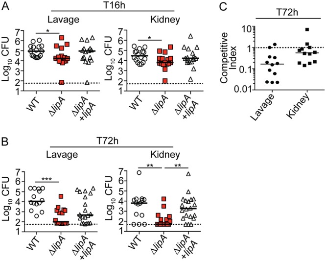
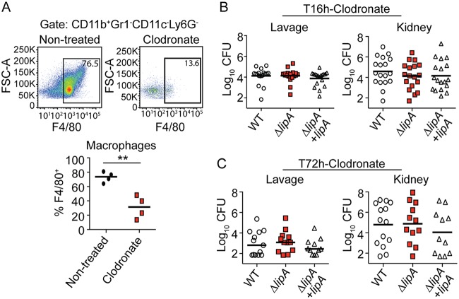
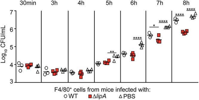
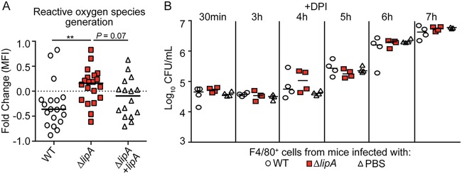
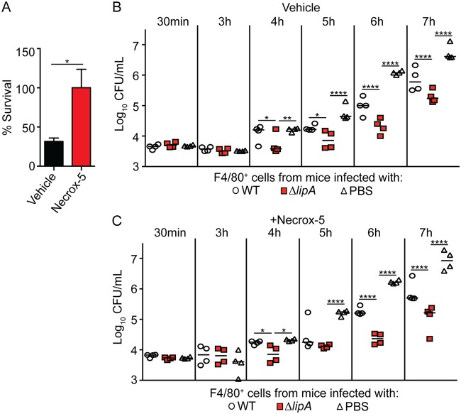
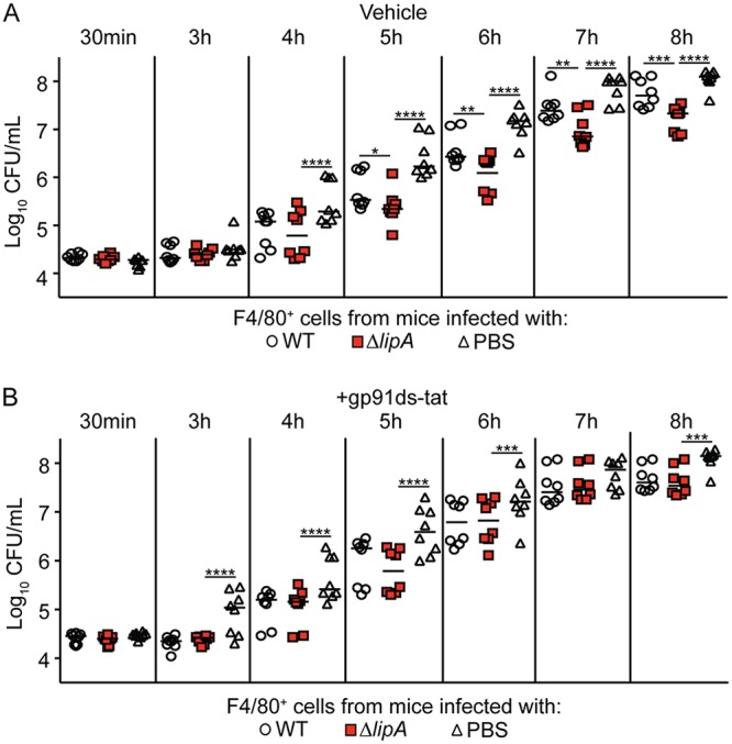
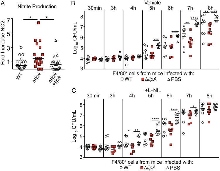
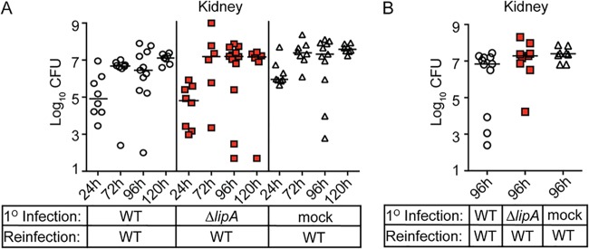
Macrophages are critical mediators of innate immunity and must be overcome for bacterial pathogens to cause disease. The Gram-positive bacterium Staphylococcus aureus produces virulence factors that impede macrophages and other immune cells. We previously determined that production of the metabolic cofactor lipoic acid by the lipoic acid synthetase, LipA, blunts macrophage activation. A ΔlipA mutant was attenuated during infection and was more readily cleared from the host. We hypothesized that bacterial lipoic acid synthesis perturbs macrophage antimicrobial functions and therefore hinders the clearance of S. aureus Here, we found that enhanced innate immune cell activation after infection with a ΔlipA mutant was central to attenuation in vivo, whereas a growth defect imparted by the lipA mutation made a negligible contribution to overall clearance. Macrophages recruited to the site of infection with the ΔlipA mutant produced larger amounts of bactericidal reactive oxygen species (ROS) and reactive nitrogen species (RNS) than those recruited to the site of infection with the wild-type strain or the mutant strain complemented with lipA ROS derived from the NADPH phagocyte oxidase complex and RNS derived from the inducible nitric oxide synthetase, but not mitochondrial ROS, were critical for the restriction of bacterial growth under these conditions. Despite enhanced antimicrobial immunity upon primary infection with the ΔlipA mutant, we found that the host failed to mount an improved recall response to secondary infection. Our data suggest that lipoic acid synthesis in S. aureus promotes bacterial persistence during infection through limitation of ROS and RNS generation by macrophages. Broadly, this work furthers our understanding of the intersections between bacterial metabolism and immune responses to infection.

Keywords: Staphylococcus aureus; lipoic acid; macrophages; metabolism; reactive nitrogen species; reactive oxygen species; virulence.


Figures

FIG 1
FIG 1
The ΔlipA mutant is attenuated during systemic infection. Bacterial burden (log10 number of CFU) in the peritoneal cavity and kidneys of mice at 16 h (T16h) after intraperitoneal infection with the WT (n = 16), ΔlipA (n = 16), or ΔlipA + lipA (n = 16) strain (A) or 72 h (T72h) after intraperitoneal infection with the WT (n = 14), ΔlipA (n = 16), or ΔlipA + lipA (n = 20) strain (B). Bars represent means. P values were determined by the Kruskal-Wallis test with Dunn’s posttest. *, P < 0.05; **, P < 0.01; ***, P < 0.001. Dashed line, limit of detection. (C) Competitive index for the lipA::erm and WT strains after coinfection (n = 12) with a 50/50 mix of the WT and lipA::erm strains for 72 h. Dashed line, competitive index of 1.
FIG 2
FIG 2
The ΔlipA mutant is not attenuated in macrophage-depleted mice. (A) Abundance of macrophages (CD11b+ Gr1 CD11c Ly6G F4/80+) in the peritoneal cavity of clodronate-treated mice or nontreated mice at 72 h after intraperitoneal infection with WT S. aureus. Representative flow cytometry plots are shown along with composite data from four clodronate-treated mice and four nontreated mice. Bars represent the median. Data were analyzed using an unpaired, two-tailed Student's t test. **, P < 0.01. FSC-A, forward scatter area. (B and C) Bacterial burden (log10 number of CFU) in the peritoneal cavity and kidneys of clodronate-treated mice at 16 h after intraperitoneal infection with the WT (n = 18), ΔlipA (n = 18), or ΔlipA + lipA (n = 18) strain (B) or 72 h after intraperitoneal infection with the WT (n = 13), ΔlipA (n = 12), or ΔlipA + lipA (n = 11) strain (C). Bars represent means. Comparisons between groups in panels B and C were not statistically significant, as determined by the Kruskal-Wallis test with Dunn’s posttest.
FIG 3
FIG 3
Macrophages recruited to the site of ΔlipA mutant infection restrict S. aureus outgrowth. Outgrowth (log10 number of CFU per milliliter) of WT S. aureus after infection of F4/80+ cells isolated from mice at 72 h after intraperitoneal infection with the WT strain (n = 4), ΔlipA mutant (n = 4), or PBS (n = 4). Bars represent medians. P values were determined by 2-way ANOVA with Tukey’s posttest. *, P < 0.05; **, P < 0.01; ****, P < 0.0001. The data shown are from one of at least three independent experiments.
FIG 4
FIG 4
Macrophages from ΔlipA mutant-infected mice produce larger amounts of ROS that control bacterial outgrowth than macrophages from mice infected with the WT or ΔlipA + lipA strain. (A) F4/80+ macrophages were sorted from immune cells harvested from the peritoneal cavities of mice at 72 h after intraperitoneal infection with the WT (n = 19), ΔlipA (n = 20), or ΔlipA + lipA (n = 16) strain and infected ex vivo with WT S. aureus at an MOI of 0.1. Macrophages were stained with the ROS indicator CellROX deep red and analyzed by flow cytometry. Fold changes in the geometric means of CellROX fluorescence in infected cells compared to those in uninfected F4/80+ cells were quantified. **, P < 0.01 by the Kruskal-Wallis test with Dunn’s posttest. The dashed line is at a value of zero, and bars represent the median. (B) Outgrowth (log10 number of CFU per milliliter) of WT S. aureus after infection of F4/80+ cells isolated from mice at 72 h after intraperitoneal infection with the WT strain (n = 4), the ΔlipA mutant (n = 4), or PBS (n = 4) and treated with the ROS inhibitor DPI. Bars represent medians. The data shown are from one of at least three independent experiments.
FIG 5
FIG 5
Macrophages isolated from ΔlipA mutant-infected mice do not use mROS to restrict bacterial outgrowth. (A) Percent survival of WT S. aureus 8 h after infecting F4/80+ cells isolated from mice treated with vehicle control (DMSO) or the mROS inhibitor Necrox-5. Error bars represent SEM (n = 8). Data were analyzed using an unpaired, two-tailed Student's t test. *, P < 0.05. (B and C) Outgrowth (log10 number of CFU per milliliter) of WT S. aureus after infection of F4/80+ cells isolated from mice at 72 h after intraperitoneal infection with the WT strain (n = 4), the ΔlipA mutant (n = 4), or PBS (n = 4) and treated with the vehicle control (DMSO) (B) or Necrox-5 (C). Bars represent medians. P values were determined by 2-way ANOVA with Tukey’s posttest. *, P < 0.05; **, P < 0.01; ****, P < 0.0001. The data shown are from one of at least two independent experiments.
FIG 6
FIG 6
NADPH oxidase-derived ROS contributes to improved control of bacterial outgrowth by macrophages isolated from ΔlipA mutant-infected mice. The outgrowth (log10 number of CFU per milliliter) of WT S. aureus after infection of F4/80+ cells isolated from mice at 72 h after intraperitoneal infection with the WT strain (n = 8), the ΔlipA mutant (n = 8), or PBS (n = 8) amd treated with the vehicle control (water) (A) or the NADPH oxidase inhibitor gp91ds-tat (B) is shown. Bars represent medians. P values were determined by 2-way ANOVA with Tukey’s posttest. *, P < 0.05; **, P < 0.01; ***, P < 0.001; ****, P < 0.0001.
FIG 7
FIG 7
RNS are important for restriction of bacterial growth by macrophages isolated from ΔlipA mutant-infected mice. F4/80+ macrophages were sorted from immune cells harvested from the peritoneal cavities of mice at 72 h after intraperitoneal infection with the WT (n = 20), ΔlipA (n = 19), or ΔlipA + lipA (n = 20) strain and stimulated overnight ex vivo with heat-killed WT S. aureus at an MOI of 10. The levels of nitrite, a breakdown of nitric oxide production, were measured by the Griess test (A). The fold induction of nitric oxide production was determined by comparing the levels of nitrite produced by infected cells to the levels produced by uninfected F4/80+ cells. *, P < 0.05 by the Kruskal-Wallis test with Dunn’s posttest. Bars represent the median. (B and C) Outgrowth (log10 number of CFU per milliliter) of WT S. aureus after infection of F4/80+ cells isolated from mice at 72 h after intraperitoneal infection with the WT strain (n = 8), the ΔlipA mutant (n = 8), or PBS (n = 8) and treated with the vehicle control (water) (B) or the iNOS inhibitor L-NIL (C). Bars represent medians. P values were determined by 2-way ANOVA with Tukey’s posttest. *, P < 0.05; **, P < 0.01; ***, P < 0.001; ****, P < 0.0001.
FIG 8
FIG 8
Immunization of mice with the ΔlipA mutant does not confer protection from secondary challenge. Mice were immunized by intraperitoneal injection with 1 × 108 CFU of the S. aureus WT and ΔlipA mutant or sterile PBS (mock immunized). At either 7 (A) or 14 (B) days after immunization, the mice were rechallenged via injection of 1 × 107 CFU of WT S. aureus into the retro-orbital sinus. The bacterial burden (log10 number of CFU) in the kidneys of mice was assessed 24 h (WT, n = 8; ΔlipA mutant, n = 8; PBS, n = 8), 72 h (WT, n = 8; ΔlipA mutant, n = 6; PBS, n = 8), 96 h (WT, n = 11; ΔlipA mutant, n = 12; PBS, n = 11), and 120 h (WT, n = 7; ΔlipA mutant, n = 6; PBS, n = 8) (A) or 96 h (WT, n = 11; ΔlipA mutant, n = 9; PBS, n = 8) (B) after secondary challenge. Bars represent medians. No statistical significance was achieved when comparing WT- and ΔlipA mutant-immunized animals.

References

    1. Spaan AN, Surewaard BG, Nijland R, van Strijp JA. 2013. Neutrophils versus Staphylococcus aureus: a biological tug of war. Annu Rev Microbiol 67:629–650. doi:10.1146/annurev-micro-092412-155746. - DOI - PubMed
    1. Rooijakkers SHM, van Kessel KPM, van Strijp J. 2005. Staphylococcal innate immune evasion. Trends Microbiol 13:596–601. doi:10.1016/j.tim.2005.10.002. - DOI - PubMed
    1. Foster TJ. 2005. Immune evasion by staphylococci. Nat Rev Microbiol 3:948–958. doi:10.1038/nrmicro1289. - DOI - PubMed
    1. Thammavongsa V, Kim HK, Missiakas D, Schneewind O. 2015. Staphylococcal manipulation of host immune responses. Nat Rev Microbiol 13:529–543. doi:10.1038/nrmicro3521. - DOI - PMC - PubMed
    1. Kourtis AP, Hatfield K, Baggs J, Mu Y, See I, Epson E, Nadle J, Kainer MA, Dumyati G, Petit S, Ray SM, Ham D, Capers C, Ewing H, Coffin N, McDonald LC, Jernigan J, Cardo D. 2019. Vital signs: epidemiology, and recent trends in methicillin-resistant and in methicillin-susceptible Staphylococcus aureus bloodstream infections—United States. MMWR Morb Mortal Wkly Rep 68:214–219. doi:10.15585/mmwr.mm6809e1. - DOI - PMC - PubMed

Publication types

MeSH terms