. 2019 Sep 19;87(10):e00278-19.
doi: 10.1128/IAI.00278-19. Print 2019 Oct.

Interference with Pseudomonas aeruginosa Quorum Sensing and Virulence by the Mycobacterial Pseudomonas Quinolone Signal Dioxygenase AqdC in Combination with the N-Acylhomoserine Lactone Lactonase QsdA

Affiliations

Interference with Pseudomonas aeruginosa Quorum Sensing and Virulence by the Mycobacterial Pseudomonas Quinolone Signal Dioxygenase AqdC in Combination with the N-Acylhomoserine Lactone Lactonase QsdA

Franziska S Birmes et al. Infect Immun. .

Abstract

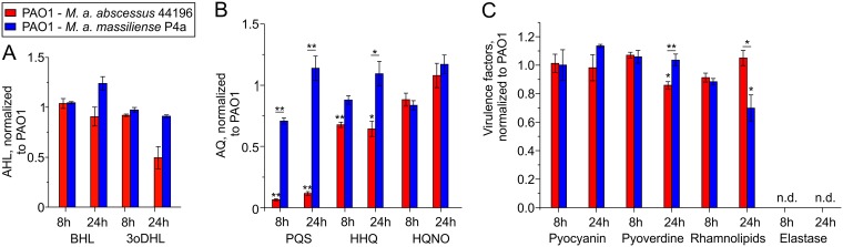
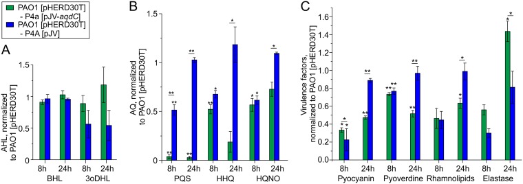
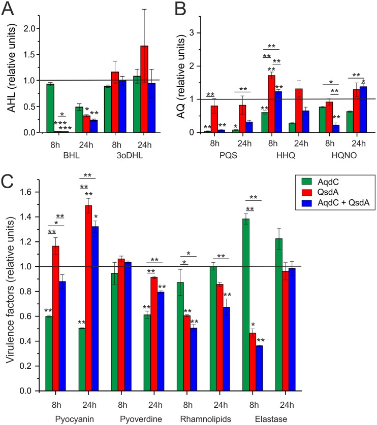
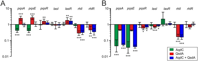
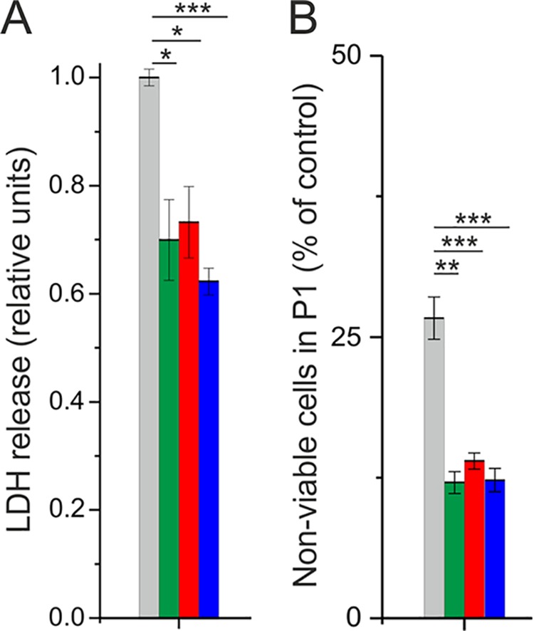
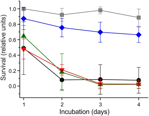
The nosocomial pathogen Pseudomonas aeruginosa regulates its virulence via a complex quorum sensing network, which, besides N-acylhomoserine lactones, includes the alkylquinolone signal molecules 2-heptyl-3-hydroxy-4(1H)-quinolone (Pseudomonas quinolone signal [PQS]) and 2-heptyl-4(1H)-quinolone (HHQ). Mycobacteroides abscessus subsp. abscessus, an emerging pathogen, is capable of degrading the PQS and also HHQ. Here, we show that although M. abscessus subsp. abscessus reduced PQS levels in coculture with P. aeruginosa PAO1, this did not suffice for quenching the production of the virulence factors pyocyanin, pyoverdine, and rhamnolipids. However, the levels of these virulence factors were reduced in cocultures of P. aeruginosa PAO1 with recombinant M. abscessus subsp. massiliense overexpressing the PQS dioxygenase gene aqdC of M. abscessus subsp. abscessus, corroborating the potential of AqdC as a quorum quenching enzyme. When added extracellularly to P. aeruginosa cultures, AqdC quenched alkylquinolone and pyocyanin production but induced an increase in elastase levels. When supplementing P. aeruginosa cultures with QsdA, an enzyme from Rhodococcus erythropolis which inactivates N-acylhomoserine lactone signals, rhamnolipid and elastase levels were quenched, but HHQ and pyocyanin synthesis was promoted. Thus, single quorum quenching enzymes, targeting individual circuits within a complex quorum sensing network, may also elicit undesirable regulatory effects. Supernatants of P. aeruginosa cultures grown in the presence of AqdC, QsdA, or both enzymes were less cytotoxic to human epithelial lung cells than supernatants of untreated cultures. Furthermore, the combination of both aqdC and qsdA in P. aeruginosa resulted in a decline of Caenorhabditis elegans mortality under P. aeruginosa exposure.

Keywords: Mycobacteroides abscessus; Pseudomonas aeruginosa; Pseudomonas quinolone signal; dioxygenase; lactonase; quorum quenching enzyme; quorum sensing.


Figures

FIG 1
FIG 1
AHLs (A), AQs (B), and virulence factor production (C) in cocultures of P. aeruginosa PAO1 with M. abscessus subsp. abscessus DSM44196 or with M. abscessus subsp. massiliense P4a, normalized to solo cultures of P. aeruginosa PAO1. CFU counts after 8 h were 2.87 × 108 ± 1.91 × 107 CFU/ml for PAO1 in solo culture; 2.26 × 108 ± 1.46 × 107 and 2.11 × 107 ± 5.85 × 106 CFU/ml for PAO1 and M. abscessus DSM44196, respectively, in coculture; and 2.27 × 108 ± 1.20 × 107 and 4.9 × 107 ± 4.73 × 106 CFU/ml for PAO1 and M. abscessus subsp. massiliense P4a, respectively, in coculture. CFU counts after 24 h were 7.22 × 108 ± 4.01 × 107 CFU/ml for PAO1 in solo culture; 8.61 × 108 ± 1.12 × 107 and 3.68 × 104 ± 6.23 × 103 CFU/ml for PAO1 and M. abscessus DSM44196, respectively, in coculture; and 6.67 × 108 ± 5.09 × 107 and 5.08 × 105 ± 6.43 × 106 CFU/ml for PAO1 and M. abscessus subsp. massiliense P4a, respectively, in coculture. Data represent means ± standard errors (SE) from three independent biological replicates. Statistical analysis was carried out using analysis of variance (ANOVA) or a Tukey honestly significant difference (HSD) test. P values assigned to individual bars refer to solo cultures of P. aeruginosa PAO1 (*, P < 0.05; **, P < 0.01).
FIG 2
FIG 2
AHLs (A), AQs (B), and virulence factor production (C) in cocultures of P. aeruginosa PAO1(pHERD30T) with M. abscessus subsp. massiliense P4a(pJV-aqdC) or with M. abscessus subsp. massiliense P4a(pJV), normalized to solo cultures of P. aeruginosa PAO1(pHERD30T). In panel A, AHL levels were normalized to the AHL concentrations in the PAO1 solo culture. CFU counts after 8 h were 4.44 × 107 ± 2.94 × 106 CFU/ml for PAO1 in solo culture; 2.77 × 107 ± 1.45 × 106 and 4.4 × 107 ± 2.2 × 106 CFU/ml for PAO1 and M. abscessus subsp. massiliense P4a(pJV-aqdC), respectively, in coculture; and 4.84 × 107 ± 1.39 × 106 and 2.37 × 107 ± 2.47 × 106 CFU/ml for PAO1 and M. abscessus subsp. massiliense P4a(pJV), respectively, in coculture. CFU counts after 24 h were 8.33 × 108 ± 1.07 × 108 CFU/ml for PAO1 in solo culture; 6.67 × 108 ± 3.33 × 107 and 2.43 × 105 ± 5.07 × 104 CFU/ml for PAO1 and M. abscessus subsp. massiliense P4a(pJV-aqdC), respectively, in coculture; and 1.07 × 109 ± 1.35 × 108 and 9.58 × 106 ± 8.92 × 104 CFU/ml for PAO1 and M. abscessus subsp. massiliense P4a(pJV), respectively, in coculture. Data represent means ± SE from three independent biological replicates. Statistical analysis was carried out using ANOVA or a Tukey HSD test. P values assigned to individual bars refer to solo cultures of P. aeruginosa PAO1(pHERD30T) (*, P < 0.05; **, P < 0.01).
FIG 3
FIG 3
Substrate specificity of AqdC, AqdC1, and Hod (A) and stability of the dioxygenases and the lactonase QsdA (B). (A) Activities of AqdC and AqdC1 were normalized to the activity toward PQS, whereas the activity of Hod was normalized to the activity of its natural substrate C1-PQS. (B) Residual activity of the quorum quenching enzymes after incubation for 8 and 24 h at 37°C in P. aeruginosa PAO1 cell suspensions or in LB medium, normalized to the values before incubation (set to 1). For determining QsdA activity in cell suspensions, the lactonase activity of untreated P. aeruginosa cell suspensions was subtracted from the total lactonase activity of QsdA-treated cell suspensions. Data represent means ± SE from three technical replicates.
FIG 4
FIG 4
AHLs (A), AQ concentrations (B), and virulence factor production (C) in cultures of P. aeruginosa PAO1 after addition of 0.3 U/ml (5.66 μg/ml) AqdC, 0.255 mg/ml QsdA, and the combination of both enzymes. Levels of virulence factors and signal molecules in P. aeruginosa PAO1 supplemented with buffer without any enzymes are set to 1 and are represented by the black lines. Data represent means ± SE from three independent biological replicates. Statistical analysis was carried out using ANOVA or a Tukey HSD test (*, P < 0.05; **, P < 0.01; ***, P < 0.001).
FIG 5
FIG 5
Relative transcription levels of quorum sensing-associated genes in P. aeruginosa PAO1 cultures in the presence of quorum quenching enzymes. Shown are fold changes in gene expression in response to the addition of 0.3 U/ml (5.66 μg/ml) AqdC, 0.255 mg/ml QsdA, and the combination of both enzymes to LB cultures of P. aeruginosa PAO1, normalized to the transcription of the internal control gene rpoS after 8 h (A) and 24 h (B). Transcript amounts of PAO1 cultures in the absence of quorum quenching enzymes were set to 1. Data represent means ± standard deviations (SD) from three biological replicates measured in at least technical duplicates each. Statistical analysis was carried out using ANOVA or a Tukey HSD test (*, P < 0.05; **, P < 0.01; ***, P < 0.001).
FIG 6
FIG 6
Effect of P. aeruginosa PAO1 supernatants on A549 lung epithelial cells. (A) Relative LDH release of A549 cells after incubation with the supernatant of enzymatically treated or untreated PAO1 cultures. LDH release of cells incubated with the supernatant of untreated PAO1 cultures was set to 1. (B) Number of necrotic cells (stained with propidium iodide) counted within the FACS gate that was generated on the basis of untreated control cells (P1) as a percentage of cell numbers in treated compared to untreated control samples. Gray bars represent incubation of A549 cells with the supernatant of untreated PAO1 cultures, and green, red, and blue bars indicate incubation of A549 cells with the supernatant of PAO1 cultures treated with 0.5 U/ml (8.5 μg/ml) AqdC, 0.38 mg/ml QsdA, and a combination of both, respectively. Data represent means ± SE from three independent biological replicates. Statistical analysis was done using Student’s t test (*, P < 0.05; **, P < 0.01; ***, P < 0.001).
FIG 7
FIG 7
Expression of both aqdC and qsdA increases survival of C. elegans under PAO1 exposure. Young adult C. elegans hermaphrodites were transferred onto NGM plates containing P. aeruginosa PAO1 strains, and survival was scored for four consecutive days. Black, P. aeruginosa PAO1(pME6032, pBBR1-MCS5) (empty vectors); green, PAO1(pME::aqdC, pBBR1-MCS5); red, PAO1(pME6032, pBBR::qsdA); blue, PAO1(pME::aqdC, pBBR::qsdA). As a negative control, the survival of C. elegans on E. coli OP50(pME::aqdC, pBBR::qsdA) lawns is depicted (gray). Data represent means ± SD from five plates with 10 worms each. Experiments were repeated three times. The survival rate of worms on PAO1(pME::aqdC, pBBR::qsdA) lawns was significantly higher (P < 0.05 by two-way ANOVA) than on PAO1(pME6032, pBBR1-MCS5) lawns in each biological replicate. Growth rates [OD600(t2) − OD600(t1)/t2t1] of recombinant P. aeruginosa PAO1 strains in LB medium (with appropriate antibiotics) at 22°C were as follows: 0.20 h−1 for PAO1(pME6032, pBBR1-MCS5), 0.15 h−1 for PAO1(pME::aqdC, pBBR1-MCS5), 0.19 h−1 for PAO1(pME6032, pBBR::qsdA), and 0.15 h−1 for PAO1(pME::aqdC, pBBR::qsdA).

Similar articles

Cited by

References

    1. Cystic Fibrosis Foundation. 2017. Annual data report 2016. Cystic fibrosis foundation patient registry. Cystic Fibrosis Foundation, Bethesda, MD.
    1. Schuster M, Greenberg EP. 2006. A network of networks: quorum-sensing gene regulation in Pseudomonas aeruginosa. Int J Med Microbiol 296:73–81. doi:10.1016/j.ijmm.2006.01.036. - DOI - PubMed
    1. Huse H, Whiteley M. 2011. 4-Quinolones: smart phones of the microbial world. Chem Rev 111:152–159. doi:10.1021/cr100063u. - DOI - PMC - PubMed
    1. Heeb S, Fletcher MP, Chhabra SR, Diggle SP, Williams P, Cámara M. 2011. Quinolones: from antibiotics to autoinducers. FEMS Microbiol Rev 35:247–274. doi:10.1111/j.1574-6976.2010.00247.x. - DOI - PMC - PubMed
    1. Papenfort K, Bassler BL. 2016. Quorum sensing signal-response systems in Gram-negative bacteria. Nat Rev Microbiol 14:576–588. doi:10.1038/nrmicro.2016.89. - DOI - PMC - PubMed

Publication types

MeSH terms

LinkOut - more resources