. 2019 Sep 1;203(5):1189-1197.
doi: 10.4049/jimmunol.1900398. Epub 2019 Jul 15.

Control of Tissue-Resident Invariant NKT Cells by Vitamin A Metabolites and P2X7-Mediated Cell Death

Affiliations

Control of Tissue-Resident Invariant NKT Cells by Vitamin A Metabolites and P2X7-Mediated Cell Death

Qingyang Liu et al. J Immunol. .

Abstract

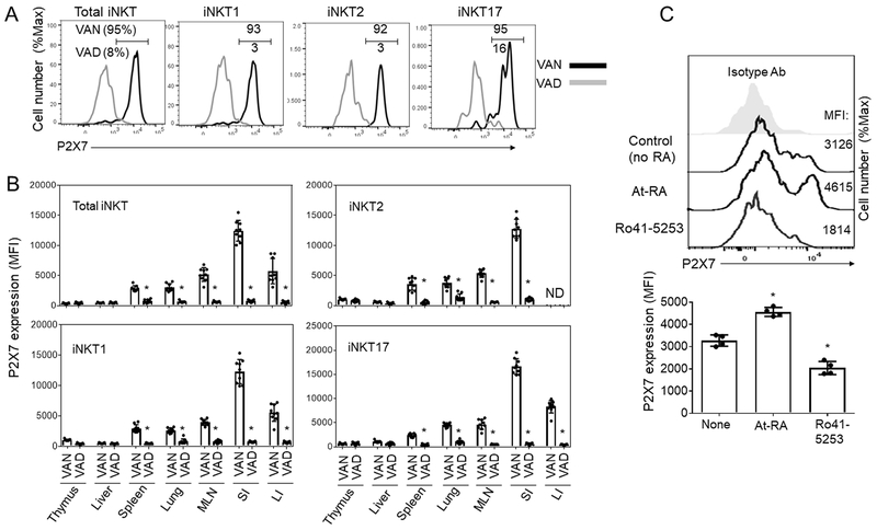
Invariant NKT (iNKT) cells provide rapid innate T cell responses to glycolipid Ags from host cells and microbes. The numbers of CD1d-restricted iNKT cells are tightly controlled in mucosal tissues, but the mechanisms have been largely unclear. We found that vitamin A is a dominant factor that controls the population size of mucosal iNKT cells in mice. This negative regulation is mediated by the induction of the purinergic receptor P2X7 on iNKT cells. The expression of P2X7 is particularly high on intestinal iNKT cells, making iNKT cells highly susceptible to P2X7-mediated cell death. In vitamin A deficiency, iNKT cells fail to express P2X7 and are, therefore, resistant to P2X7-mediated cell death, leading to iNKT cell overpopulation. This phenomenon is most prominent in the intestine. We found that iNKT cells are divided into CD69+ sphingosine-1-phosphate receptor 1 (S1P1)- tissue resident and CD69- S1P1+ nonresident iNKT cells. The CD69+ S1P1- tissue-resident iNKT cells highly express P2X7 and are effectively controlled by the P2X7 pathway. The regulation of iNKT cells by vitamin A by the P2X7 pathway is important to prevent aberrant expansion of effector cytokine-producing iNKT cells. Our findings identify a novel role of vitamin A in regulating iNKT cell homeostasis in many tissues throughout the body.


Conflict of interest statement

Disclosure: The authors declare no competing financial interests.

Figures

Fig. 1.
Fig. 1.. iNKT cell dysplasia in vitamin A deficiency and differential expression of P2X7 by iNKT cells of different tissues.
(A) Numbers of CD1d-Tetramer+ iNKT cells in the spleen and small intestine (SI) of vitamin A-deficient (VAD) versus normal (VAN) mice. Mice were fed VAN or VAD diets for 10-12 weeks from birth and examined for indicated iNKT cells. (B, C) P2X7 is highly expressed by CD1d-Tetramer+ iNKT cells in the intestines and MLN. Representative and combined data (n=6 for A; n=4 for C) are shown. All error bars indicate SEM. *Significant differences (P values <0.05) from control or between two groups.
Fig. 2.
Fig. 2.. Vitamin A is required for normal expression of P2X7 by iNKT cells.
(A) P2X7 expression by indicated CD1d-Tetramer+ iNKT cells in the intestine of VAN versus VAD mice. (B) P2X7 expression by iNKT cells of VAN versus VAD mice. (C) Impact of RARα agonist and antagonist on P2X7 expression by iNKT cells. Splenocytes were cultured in the presence of IL-2 and RA or Ro-41-5253 for 4 days, and P2X7 expression by CD1d-Tet+ iNKT cells were examined (n=4). OCH was added during the first 24 h for cell stimulation. *Significant differences (P values <0.05) from VAN or control groups.
Fig. 3.
Fig. 3.. P2X7 activation-induced death of iNKT cells in WT versus P2X7−/− mice.
(A) Ex vivo cell death of iNKT cells, isolated from the SI of WT and P2X7−/− mice, in response to NAD. Annexin V and propidium iodide (PI) staining was performed. (B) In vivo depletion of iNKT cells in the small intestine of WT and P2X7−/− mice 1 day after NAD injection. Absolute cell numbers are shown (n=4). *Significant differences (P values <0.05) from WT or control mice.
Fig. 4.
Fig. 4.. Increased numbers of iNKT cells in the intestine of P2X7−/− mice.
(A) Comparison of WT and P2X7−/− mice for iNKT cells in the small intestinal villi in the ileum. CD1d+Teramer+ cells per 10,000 μm2 of lamina propria areas were counted. Cell counts for 18 villi from 3 mice are shown for each group. (B) Numbers of iNKT cells and iNKT cell subsets in the lamina propria of SI and LI. (C, D) Numbers of iNKT cells in WT and P2X7−/− mice following ART2.2 blockade. Anti-ART2.2 single chain antibody was injected i.v. into mice 30 min before euthanasia. Combined data (n=6 for B, n=4 for D) are shown. *Significant differences (P values <0.05) from wild type control mice.
Fig. 5.
Fig. 5.. Over-production of iNKT cell-derived effector cytokines in P2X7−/− mice.
(A, B) Comparison of WT and P2X7−/− mice for iNKT cells producing indicated cytokines in the small intestinal lamina propria. Frequency of iNKT cells producing indicated cytokines in the small intestinal lamina propria is shown. Mice were injected with OCH i.g. and examined 24 hours later. (C) Cytokine concentrations in the blood plasma in WT versus P2X7−/− mice were determined. Mice were injected with OCH i.g. or i.v. and examined 24 hours later. Representative and combined data (n=4) are shown. *Significant differences (P values <0.05) between two groups.
Fig. 6.
Fig. 6.. P2X7−/− iNKT cells have greatly increased population potentials.
(A) Competitive population of WT and P2X7−/− iNKT cells in the population of indicated organs. Enriched CD1d-Tet+ cells were isolated from the spleen tissues of WT and P2X7−/− mice and were injected into Rag1-deficient mice. The mice were euthanized 3 weeks later, and iNKT cell numbers in indicated tissues were determined by flow cytometry. (B) Competitive population of WT and P2X7−/− iNKT subsets in the indicated organs. Representative and combined data (n=6) are shown.
Fig. 7.
Fig. 7.. Tissue-resident CD69+ iNKT cells highly express P2X7 and are effectively controlled by P2X7.
(A) Expression of P2X7 by CD69 and CD69+ iNKT cells. (B) Expression of S1PR1 by CD69 and CD69+ iNKT cells. (C) Replacement rates of CD69 and CD69+ iNKT cells by non-host-derived iNKT cells in indicated tissues of parabiosis mice. CD69 and CD69+ iNKT cells in parabiosis mice between congenic CD45.1 and CD45.2 WT mice. (D) Abnormal expansion of CD69+ iNKT cells in P2X7-deficient mice. (E) Comparison of the population of CD69 and CD69+ iNKT cells in the parabiosis mice between WT CD45.1 and P2X7−/− CD45.2 mice. Representative and combined data (n=3-7) are shown. *Significant differences (P values <0.05) from control groups.
Fig. 8.
Fig. 8.. Vitamin A deficiency fails to increase the numbers of iNKT cells in the absence of P2X7 expression.
Comparison of the numbers of P2X7−/− iNKT cells in vitamin A normal (VAN) and deficient (VAD) conditions. P2X7−/− mice were fed VAN or VAD diet for 11 weeks from birth and examined for indicated iNKT cells. Combined data (n=5-6) are shown. All error bars indicate SEM. *Significant differences (P values <0.05) between two groups.

Similar articles

Cited by

References

    1. Kim CH 2018. Control of Innate and Adaptive Lymphocytes by the RAR-Retinoic Acid Axis. Immune Netw 18: e1. - PMC - PubMed
    1. Mora JR, Iwata M, and von Andrian UH. 2008. Vitamin effects on the immune system: vitamins A and D take centre stage. Nat Rev Immunol 8: 685–698. - PMC - PubMed
    1. Iwata M 2009. Retinoic acid production by intestinal dendritic cells and its role in T-cell trafficking. Semin Immunol 21: 8–13. - PubMed
    1. Raverdeau M, and Mills KH. 2014. Modulation of T cell and innate immune responses by retinoic Acid. J Immunol 192: 2953–2958. - PubMed
    1. An D, Oh SF, Olszak T, Neves JF, Avci FY, Erturk-Hasdemir D, Lu X, Zeissig S, Blumberg RS, and Kasper DL. 2014. Sphingolipids from a symbiotic microbe regulate homeostasis of host intestinal natural killer T cells. Cell 156: 123–133. - PMC - PubMed

Publication types