. 2019 Sep 11;39(37):7244-7259.
doi: 10.1523/JNEUROSCI.2973-18.2019. Epub 2019 Jul 15.

Early Life Inflammation Increases CA1 Pyramidal Neuron Excitability in a Sex and Age Dependent Manner through a Chloride Homeostasis Disruption

Affiliations

Early Life Inflammation Increases CA1 Pyramidal Neuron Excitability in a Sex and Age Dependent Manner through a Chloride Homeostasis Disruption

Carlos D Gomez et al. J Neurosci. .

Abstract

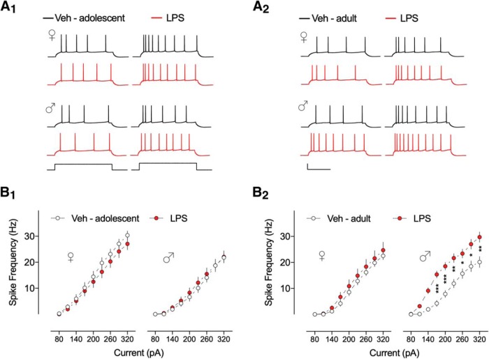
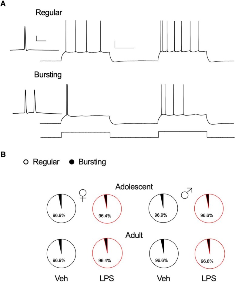
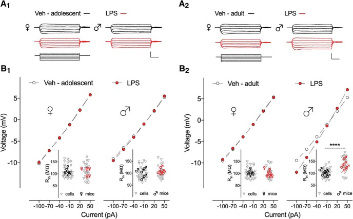
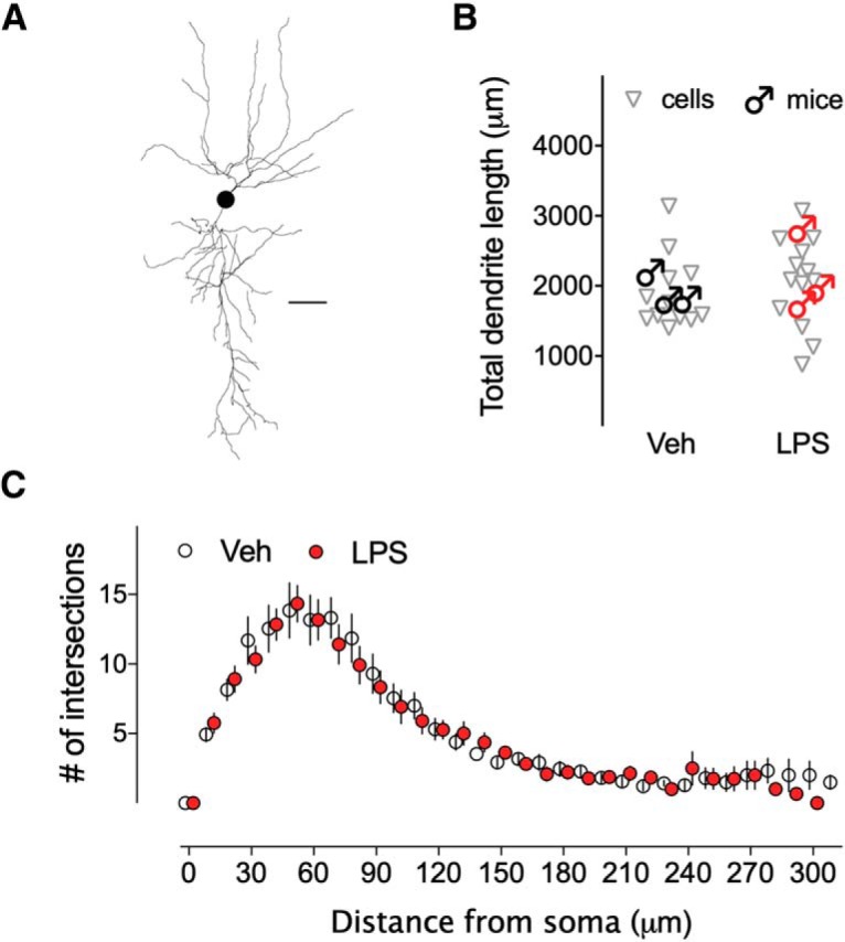
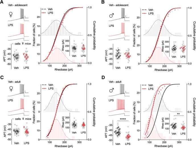
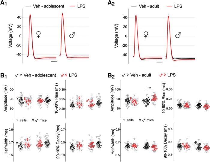
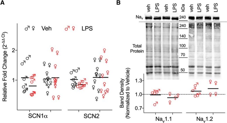
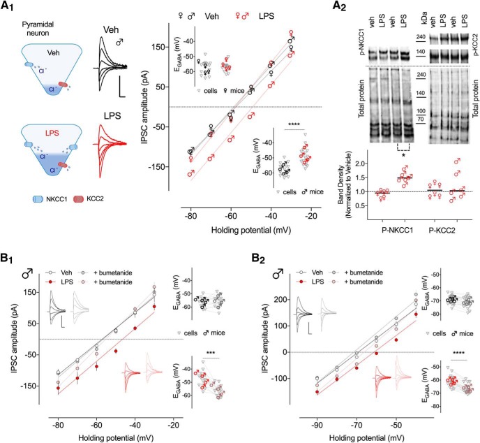
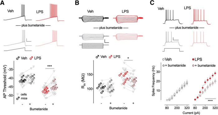
Early life, systemic inflammation causes long-lasting changes in behavior. To unmask possible mechanisms associated with this phenomenon, we asked whether the intrinsic membrane properties in hippocampal neurons were altered as a consequence of early life inflammation. C57BL/6 mice were bred in-house and both male and female pups from multiple litters were injected with lipopolysaccharide (LPS; 100 μg/kg, i.p.) or vehicle at postnatal day (P)14, and kept until adolescence (P35-P45) or adulthood (P60-P70), when brain slices were prepared for whole-cell and perforated-patch recordings from CA1 hippocampal pyramidal neurons. In neurons of adult male mice pretreated with LPS, the number of action potentials elicited by depolarizing current pulses was significantly increased compared with control neurons, concomitant with increased input resistance, and a lower action potential threshold. Although these changes were not associated with changes in relevant sodium channel expression or differences in capacitance or dendritic architecture, they were linked to a mechanism involving intracellular chloride overload, revealed through a depolarized GABA reversal potential and increased expression of the chloride transporter, NKCC1. In contrast, no significant changes were observed in neurons of adult female mice pretreated with LPS, nor in adolescent mice of either sex. These data uncover a potential mechanism involving neonatal inflammation-induced plasticity in chloride homeostasis, which may contribute to early life inflammation-induced behavioral alterations.SIGNIFICANCE STATEMENT Early life inflammation results in long-lasting changes in many aspects of adult physiology. In this paper we reveal that a brief exposure to early life peripheral inflammation with LPS increases excitability in hippocampal neurons in a sex- and age-dependent manner through a chloride homeostasis disruption. As this hyperexcitability was only seen in adult males, and not in adult females or adolescent animals of either sex, it raises the possibility of a hormonal interaction with early life inflammation. Furthermore, as neonatal inflammation is a normal feature of early life in most animals, as well as humans, these findings may be very important for the development of animal models of disease that more appropriately resemble the human condition.

Keywords: GABA reversal potential; hippocampus; inflammation; intrinsic membrane properties; lipopolysaccharide; sex differences.


Figures

Figure 1.
Figure 1.
Neonatal inflammation alters the firing frequency of hippocampal neurons from adult male mice. A, Voltage response traces from CA1 pyramidal neurons from (A1) adolescent and (A2) adult mice treated with Veh (dark line) or LPS (red line) at postnatal day 14, elicited by 500 ms duration current injections ranging from 80 to 320 pA in 30 pA increments. Calibration: 30 mV/170 pA, 200 ms. B1, Summary data show no differences in the number of APs for Veh and LPS-treated adolescent female and male mice (n = 28–32 cells, 5–6 mice/group). B2, Firing frequency of CA1 pyramidal neurons from LPS-treated adult male mice was significantly higher than Veh control group (n = 29–31 cells, 5–8 mice/group). Number of APs for adult female groups was unchanged (n = 28–32 cells, 6–7 mice/group). Error bars indicate SEM. *p < 0.05, **p < 0.01, ***p < 0.001.
Figure 2.
Figure 2.
Distribution of regular and bursting spiking pattern of hippocampal neurons. A, CA1 pyramidal neurons respond to step current injections by firing APs in two distinct patterns: regular spiking (top) or bursting (bottom). Bursting neurons display a burst of two or more APs with an instantaneous frequency >100 Hz, whereas regular spiking neurons display single spikes. However, when the current injection is increased, bursting neurons typically fire additional bursts before switching to single APs at the end of the step, and regular neurons fire single spikes with decreased interspike intervals. Insets, Magnified view of high-frequency bursts. Calibration: 20 mV/250 pA, 200 ms; inset, 20 mV/10 ms. B, Pie charts show percentages of all regular and bursting CA1 pyramidal cells recorded, and no changes in any of the groups as a function of either sex or neonatal inflammation were found.
Figure 3.
Figure 3.
Neonatal inflammation increases the input resistance of hippocampal neurons from adult male mice. A, Sample traces of voltage responses to hyperpolarizing and depolarizing current steps from −100 to 50 pA in 30 pA increments (500 ms duration) of CA1 pyramidal neurons from (A1) adolescent and (A2) adult mice treated with Veh (dark line) or LPS (red line) at P14. Calibration: 10 mV/120 pA, 100 ms. B, Current–voltage (I–V) plots for (B1) adolescent and (B2) adult mice showing mean ± SEM voltage responses to current injections as depicted in A. Significant differences between I–V curves were only observed for Veh and LPS-treated adult male mice (B2, right; p < 0.0001, n = 29–31 cells, 5–8 mice/group). B1, B2, Insets, Plots depicting Rin of individual CA1 pyramidal neurons (▿) and average values per mouse (females, ♀; males, ♂) from respective groups calculated from the slope of the current voltage plots. ****p < 0.0001.
Figure 4.
Figure 4.
General morphology of CA1 pyramidal neurons in adult male mice is unaltered by neonatal LPS. A, Three-dimensional reconstruction of a representative pyramidal neuron showing the dendritic architecture. Calibration, 30 μm. B, Scatter graph shows the total dendritic length of individual cells and average per mouse (n = 12–13 cells, 3 mice/group). C, Sholl analysis of apical dendrites of CA1 pyramidal neurons depict the number of intersections for each concentric circle, starting from the point in the center of the cell body, and analyzed per 10 μm concentric circle. Error bars indicate SEM.
Figure 5.
Figure 5.
Neonatal inflammation alters the firing threshold of hippocampal neurons from adult male but not female mice. AD, Representative traces of voltage responses to the indicated ramp current injections (2 s duration) of CA1 pyramidal neurons of adolescent and adult mice treated with Veh (dark line) or LPS (red line) at P14. Calibration: 20 mV/200 pA, 500 ms. Scatter graphs on left show AP thresholds (APTs) of individual cells and average values per mouse (n = 26–31 cells, 5–8 mice/group). The fractional distribution of cells firing first AP relative to rheobase (depicted as vertical bars and smoothed line on right) are overlaid by their respective cumulative probabilities. Insets, Rheobase plotted for individual cells and average values per mouse (n = 26–31 cells, 5–8 mice/group). **p < 0.01, ****p < 0.0001.
Figure 6.
Figure 6.
Neonatal inflammation does not affect AP kinetics of CA1 hippocampal neurons from adolescent or adult mice. A, AP waveforms for CA1 pyramidal neurons from (A1) adolescent and (A2) adult mice, are depicted as average voltage response of the first AP elicited by ramp current injections from the total traces per each group (Veh, dark line; LPS, red line). Calibration, 2 ms. B1, B2, Summary scatter graphs plotting individual cells and average values per mouse (n = 26–31 cells, 5–8 mice/group) for AP amplitude, half-width, 10–90% rise time, and 90–10% decay time. Only AP amplitude for adult male LPS pretreated mice was changed. **p < 0.01.
Figure 7.
Figure 7.
Neonatal inflammation did not modify the mRNA and protein expression of voltage gated sodium channel subunits in the hippocampus of adult mice. A, Summary scatter graph of mean (horizontal line) and individual animal (6–10 mice/group) relative expression of SCN1α and SCN2 (normalized to vehicle, 2−ΔΔCt) in adult male and female mice treated with LPS or vehicle on P14. B, Top, Immunoblots of Nav1.1 and 1.2 (∼240 kDa) from hippocampal membranes in control and neonatally inflamed adult mice. Quantification of Nav1.1 and 1.2 band-density measurements were normalized to the corresponding vehicle whole-lane total protein of the same membrane (lower gel, Total protein) using Image Lab 6.0 software. No significant change in Nav1.1 and 1.2 protein levels was detected as a result of the LPS treatment (N = 4 mice/group; each symbol represents 1 animal and the horizontal lines are the mean).
Figure 8.
Figure 8.
Whole-cell and gramicidin perforated-patch recordings show that EGABA is depolarized in hippocampal CA1 pyramidal neurons of neonatally inflamed male mice. A1, Left, Top diagram illustrating chloride homeostasis in a vehicle-treated adult pyramidal cell; low relative activity of NKCC1 and high activity of KCC2 maintains low intracellular chloride (blue fill). Bottom diagram illustrates chloride homeostasis in an adult neonatally inflamed pyramidal cell with a more active (larger blue barrel) NKCC1 and normal activity KCC2 (red barrel) maintaining a higher intracellular chloride level (blue fill). Right, whole-cell, representative evoked IPSC recordings of CA1 pyramidal neurons from adult male control (dark lines) and neonatally inflamed (red lines) mice. Calibration: 100 pA, 20 ms. Whole-cell, Current–voltage (I–V) curve of evoked IPSCs recorded at different holding potentials from −80 to −30 mV in 10 mV increments of 15–19 hippocampal neurons from control (black) or inflamed (red) mice. Inset, Plot shows individual cell EGABA values and average values per mouse obtained from all I–V curves indicating a depolarizing shift in inflamed male, but not female mice (n = 15–19 cells, 4 mice/group). A2, Top, Immunoblots of p-NKCC1 (∼140 kDa) and p-KCC2 (∼140 and ∼270 kDa) from CA1 hippocampal tissue in control and neonatally inflamed adult mice. Quantification of p-NKCC1 and p-KCC2 band density measurements were normalized to the corresponding vehicle whole-lane total protein of the same membrane (lower gel, Total protein) using Image Lab 6.0 software. Significant increase in p-NKCC1 protein level was found only in adult male mice previously exposed to LPS at P14 (N = 6 mice/group; each symbol represents one animal and the horizontal lines are the mean). Whole-cell (B1) and gramicidin (B2) recordings show that acutely applied bumetanide (10 μm) is without effect on EGABA in vehicle-treated animals (control, black; bumetanide, gray). Insets, Representative IPSC recordings and EGABA of individual cells and average values per mouse in the two groups. Interestingly, acutely applied bumetanide (10 μm; LPS, red; bumetanide, pink) reversed the neonatal inflammation-induced depolarizing shift in EGABA; insets show representative IPSCs and individual EGABA of individual cells and average values per mouse in the two groups (n = 12–20 cells, 4 mice/group). Error bars indicate SEM. *p < 0.05, ***p = 0.0001, ****p < 0.0001.
Figure 9.
Figure 9.
Inhibition of chloride overload with acutely applied bumetanide resets the AP threshold and Rin, and reverses the hyperexcitability in adult neonatally inflamed CA1 pyramidal cells. A, Representative traces of voltage responses to the indicated ramp current injections before (top traces) and after (bottom traces) bath application of bumetanide (10 μm) from CA1 pyramidal neurons of adult male mice treated with Veh (black/gray) or LPS (red/pink) at P14. Calibration: 20 mV/200 pA, 500 ms. Bottom summary scatter graphs of individual cells and average per mouse show that bumetanide restored the atypical hyperpolarized AP threshold induced by neonatal inflammation without any change in controls. B, Bumetanide also reversed the neonatal inflammation-induced increased in Rin (colors as in A). Top, Representative voltage responses to current injections. Calibration: 10 mV/100 pA, 100 ms. Lower individual cellular Rin and average per mouse before and after bumetanide in Veh and LPS pretreated groups. C, Bumetanide reduced firing frequency (top, representative traces; calibration: 20 mV/170 pA, 200 ms; bottom, plots of firing frequency in response to current injections; colors as in A), in neonatally inflamed (LPS) cells, without any significant effect in hippocampal neurons in control group. n = 15–17 cells, 5 mice/group for each dataset. *p < 0.05, ***p < 0.001, ****p < 0.0001.

Similar articles

Cited by

References

    1. Acharjee S, Verbeek M, Gomez CD, Bisht K, Lee B, Benoit L, Sharkey KA, Benediktsson A, Tremblay ME, Pittman QJ (2018) Reduced microglial activity and enhanced glutamate transmission in the basolateral amygdala in early CNS autoimmunity. J Neurosci 38:9019–9033. 10.1523/JNEUROSCI.0398-18.2018 - DOI - PMC - PubMed
    1. Alfonsa H, Merricks EM, Codadu NK, Cunningham MO, Deisseroth K, Racca C, Trevelyan AJ (2015) The contribution of raised intraneuronal chloride to epileptic network activity. J Neurosci 35:7715–7726. 10.1523/JNEUROSCI.4105-14.2015 - DOI - PMC - PubMed
    1. Barmashenko G, Hefft S, Aertsen A, Kirschstein T, Köhling R (2011) Positive shifts of the GABAA receptor reversal potential due to altered chloride homeostasis is widespread after status epilepticus. Epilepsia 52:1570–1578. 10.1111/j.1528-1167.2011.03247.x - DOI - PubMed
    1. Ben-Ari Y. (2006) Seizures beget seizures: the quest for GABA as a key player. Crit Rev Neurobiol 18:135–144. 10.1615/CritRevNeurobiol.v18.i1-2.140 - DOI - PubMed
    1. Ben-Ari Y. (2017) NKCC1 chloride importer antagonists attenuate many neurological and psychiatric disorders. Trends Neurosci 40:536–554. 10.1016/j.tins.2017.07.001 - DOI - PubMed

Publication types

MeSH terms

Substances

Grants and funding