Review
. 2019 Nov 1;11(11):a035485.
doi: 10.1101/cshperspect.a035485.

Ca2+ Signaling and Regeneration

Affiliations
Review

Ca2+ Signaling and Regeneration

Jonathan S Marchant. Cold Spring Harb Perspect Biol. .

Abstract

Regeneration is the process by which lost or damaged tissue is replaced in adult organisms. Some organisms exhibit robust regenerative capabilities, while others, including humans, do not. Understanding the molecular principles governing the regenerative malleability of different organisms is of fundamental biological interest. Further, this problem has clear impact for the field of "regenerative medicine," which aspires to understand how human cells, tissues, and organs may be restored to normal function in scenarios of disease, damage, or age-related decline. This review will focus on the planarian flatworm as a powerful model system for studying the role of Ca2+ signals in regeneration. These invertebrate animals display an astounding innate regenerative capacity capable of regenerating complete organisms from tiny, excised fragments. New knowledge and methodological capabilities in this system highlight the potential for studying the role of Ca2+ signaling at multiple stages of the regenerative blueprint that controls stem cell behavior in vivo.


Figures

Figure 1.
Figure 1.
Planarian morphology and classification. (A, Top), morphology of the planarian Dugesia japonica. Flatworms are bilaterally symmetrical and dorsoventrally flattened, displaying a clear head (anterior, “a”)-to-tail (posterior, “p”) polarity. (Bottom) Examples of D. japonica polarity defects after regeneration of trunk fragments (region illustrated by box in A) following RNAi of key patterning genes β-catenin-1 (yielding two-headed animals, eyespots shown by red arrows, and pharynx in green) or APC-1 (yielding two-tailed animals). (B) Systematic classification of free-living planaria and parasitic worms within the phylum Platyhelminthes.
Figure 2.
Figure 2.
The diverse planarian ionotropic toolkit. Cladogram describing the Dugesia japonica voltage-gated-like ion channel superfamily. Dashed groupings indicated predicted gene loss in the parasitic flatworm Schistosoma mansoni. Channel topology is shown adjacent to the predicted groupings. Prediction of sequence number is intentionally conservative, encompassing only sequences with conservation of key motifs and architecture with human counterparts. Accessory ion channel subunits (nonpore forming) are not included in this figure. Sequence analysis supporting attribution and alignment is provided in Chan et al. (2017b), from which the figure has been modified.
Figure 3.
Figure 3.
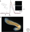
Praziquantel (PZQ) evoked bipolarity and Ca2+ signaling. (A) PZQ-evoked Ca2+ signals in dissociated planarian cells. (Inset) Structure of PZQ. Colored traces show fluo-4 fluorescence traces in dissociated single planarian cells on addition of vehicle (DMSO) and PZQ (100 µm). (Right) Images of fluorescence from cells at timepoints “1” and “2” during confocal imaging trace. (Figure modified from data in Zhang et al. 2011.) (B) Bipolar planarian produced by PZQ treatment of trunk fragment.

References

    1. Accorsi A, Williams MM, Ross EJ, Robb SMC, Elliott SA, Tu KC, Alvarado AS. 2017. Hands-on classroom activities for exploring regeneration and stem cell biology with planarians. Am Biol Teach 79: 208–223. 10.1525/abt.2017.79.3.208 - DOI
    1. Agata K, Soejima Y, Kato K, Kobayashi C, Umesono Y, Watanabe K. 1998. Structure of the planarian central nervous system (CNS) revealed by neuronal cell markers. Zoolog Sci 15: 433–440. 10.2108/zsj.15.433 - DOI - PubMed
    1. An Y, Kawaguchi A, Zhao C, Toyoda A, Sharifi-Zarchi A, Mousavi SA, Bagherzadeh R, Inoue T, Ogino H, Fujiyama A, et al. 2018. Draft genome of Dugesia japonica provides insights into conserved regulatory elements of the brain restriction gene nou-darake in planarians. Zoological Lett 4: 24 10.1186/s40851-018-0102-2 - DOI - PMC - PubMed
    1. Arenas OM, Zaharieva EE, Para A, Vásquez-Doorman C, Petersen CP, Gallio M. 2017. Activation of planarian TRPA1 by reactive oxygen species reveals a conserved mechanism for animal nociception. Nat Neurosci 20: 1686–1693. 10.1038/s41593-017-0005-0 - DOI - PMC - PubMed
    1. Beane WS, Morokuma J, Adams DS, Levin M. 2011. A chemical genetics approach reveals H,K-ATPase-mediated membrane voltage is required for planarian head regeneration. Chem Biol 18: 77–89. 10.1016/j.chembiol.2010.11.012 - DOI - PMC - PubMed

Publication types

LinkOut - more resources