

Linear magnetic resonance imaging measurements of the hippocampal formation differ in young versus old dogs
- PMID: 31308154
- PMCID: PMC6817983
- DOI: 10.1136/vr.105243
Linear magnetic resonance imaging measurements of the hippocampal formation differ in young versus old dogs
Abstract
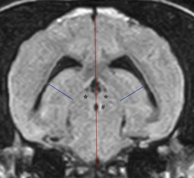
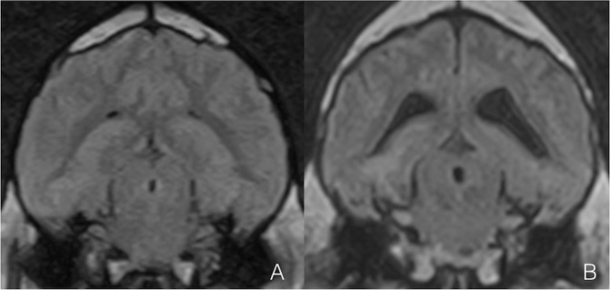
Age-related hippocampal formation (HF) atrophy has been documented on MRI studies using volumetric analysis and visual rating scales.This retrospective cross-sectional study aimed to compare linear MRI measurements of the HF between young (1-3 years) and old (>10 years) non-brachycephalic dogs, with normal brain anatomy and cerebrospinal fluid (CSF) analysis. Right and left hippocampal formation height (HFH), height of the brain (HB) and mean HFH/HB ratio were measured by two observers on a transverse T2 fluid-attenuated inversion recovery sequence containing rostral colliculi and mesencephalic aqueduct.119 MRI studies were enrolled: 75 young and 44 old dogs. Left and right HFH were greater (p<0.0001) in young, while HB was greater in old dogs (p=0.024). Mean HFH/HB ratio was 15.66 per cent and 18.30 per cent in old and young dogs (p<0.0001). No differences were found comparing measurements between epileptic and non-epileptic dogs.Old dogs have a greater HB; this may represent the different study populations or a statistical phenomenon. Ageing affects HF linear measurements. A reduction of mean HFH/HB ratio between 18.30 per cent and 15.66 per cent should be considered a physiological age-related process of the canine lifespan. The use of mean HFH/HB ratio could be considered for quantifying brain atrophy in elderly dogs.
Keywords: age-related changes; brain; dog; hippocampal formation; hippocampus; magnetic resonance imaging (mri).
© British Veterinary Association 2019. Re-use permitted under CC BY-NC. No commercial re-use. Published by BMJ.
Conflict of interest statement
Competing interests: None declared.
Figures
Similar articles
-
Canine cognitive dysfunction patients have reduced total hippocampal volume compared with aging control dogs: A comparative magnetic resonance imaging study.Open Vet J. 2021 Jan;10(4):438-442. doi: 10.4314/ovj.v10i4.11. Epub 2020 Dec 1. Open Vet J. 2021. PMID: 33614439 Free PMC article.
-
Evaluation of interthalamic adhesion size as an indicator of brain atrophy in dogs with and without cognitive dysfunction.Vet Radiol Ultrasound. 2017 Sep;58(5):581-587. doi: 10.1111/vru.12528. Epub 2017 Jul 13. Vet Radiol Ultrasound. 2017. PMID: 28707338
-
Hippocampal size did not differ between epileptic and non-epileptic dogs using volumetric and subjective methods.Acta Vet Hung. 2021 Apr 8;69(1):1-8. doi: 10.1556/004.2021.00006. Acta Vet Hung. 2021. PMID: 33835940
-
Clinical magnetic resonance volumetry of the hippocampus in 58 epileptic dogs.Vet Radiol Ultrasound. 2010 Sep-Oct;51(5):485-90. doi: 10.1111/j.1740-8261.2010.01700.x. Vet Radiol Ultrasound. 2010. PMID: 20973379
-
Comparison of magnetic resonance imaging sequences in dogs with multi-focal intracranial disease.J Small Anim Pract. 2008 Dec;49(12):634-40. doi: 10.1111/j.1748-5827.2008.00628.x. Epub 2008 Aug 5. J Small Anim Pract. 2008. PMID: 18684139
Cited by
-
Canine cognitive dysfunction patients have reduced total hippocampal volume compared with aging control dogs: A comparative magnetic resonance imaging study.Open Vet J. 2021 Jan;10(4):438-442. doi: 10.4314/ovj.v10i4.11. Epub 2020 Dec 1. Open Vet J. 2021. PMID: 33614439 Free PMC article.
-
Delineation of the Feline Hippocampal Formation: A Comparison of Magnetic Resonance Images With Anatomic Slices.Front Vet Sci. 2019 Nov 8;6:358. doi: 10.3389/fvets.2019.00358. eCollection 2019. Front Vet Sci. 2019. PMID: 31781578 Free PMC article.
-
Age-related brain atrophy in cats without apparent neurological and behavioral signs using voxel-based morphometry.Front Vet Sci. 2022 Nov 24;9:1071002. doi: 10.3389/fvets.2022.1071002. eCollection 2022. Front Vet Sci. 2022. PMID: 36504872 Free PMC article.
-
Imbalance in the ratio between mineralocorticoid and glucocorticoid receptors and neurodegeneration in the dentate gyrus of aged dogs.Vet World. 2022 Nov;15(11):2543-2550. doi: 10.14202/vetworld.2022.2543-2550. Epub 2022 Nov 10. Vet World. 2022. PMID: 36590120 Free PMC article.
References
-
- Su MY, Head E, Brooks WM, et al. . Magnetic resonance imaging of anatomic and vascular characteristics in a canine model of human aging. Neurobiol Aging 1998;19:479–85. - PubMed
MeSH terms
LinkOut - more resources
Full Text Sources
Medical
Research Materials
Miscellaneous
