. 2019 Aug 30;294(35):13073-13092.
doi: 10.1074/jbc.RA118.007331. Epub 2019 Jul 15.

A herpesvirus transactivator and cellular POU proteins extensively regulate DNA binding of the host Notch signaling protein RBP-Jκ to the virus genome

Affiliations

A herpesvirus transactivator and cellular POU proteins extensively regulate DNA binding of the host Notch signaling protein RBP-Jκ to the virus genome

Olga Gonzalez-Lopez et al. J Biol Chem. .

Abstract

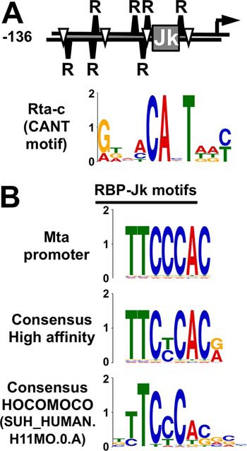
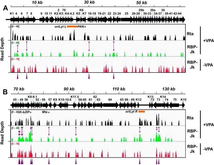
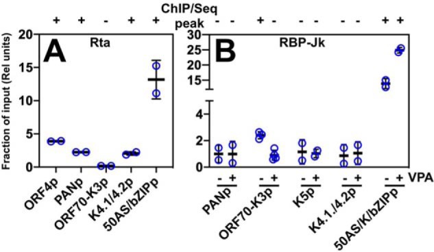
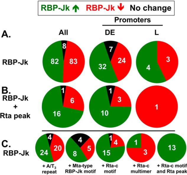
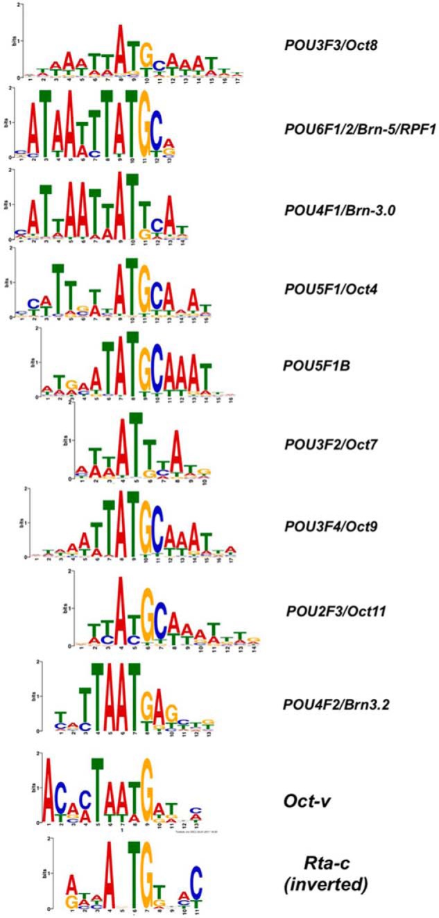
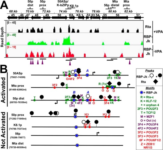
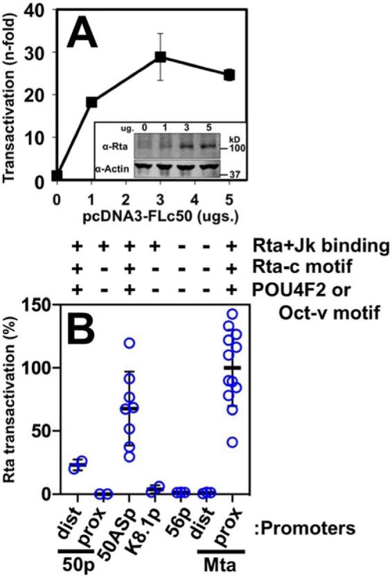
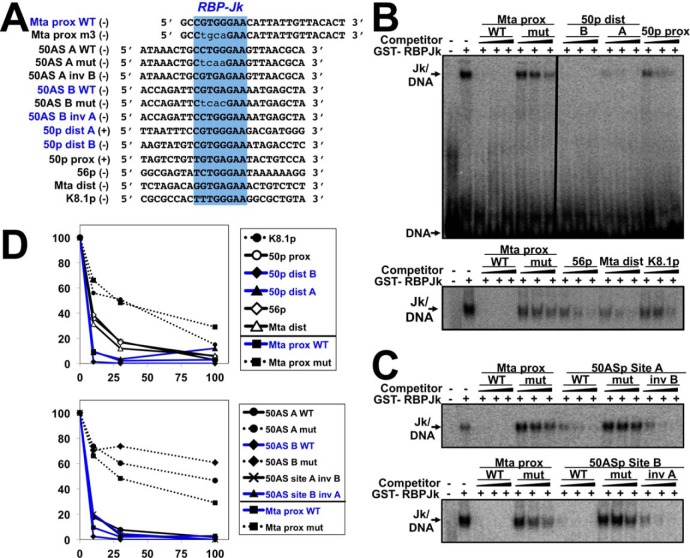
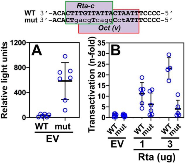
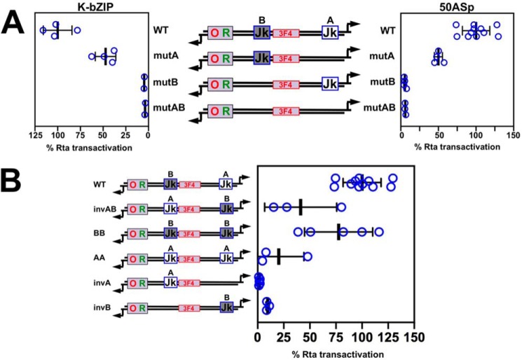
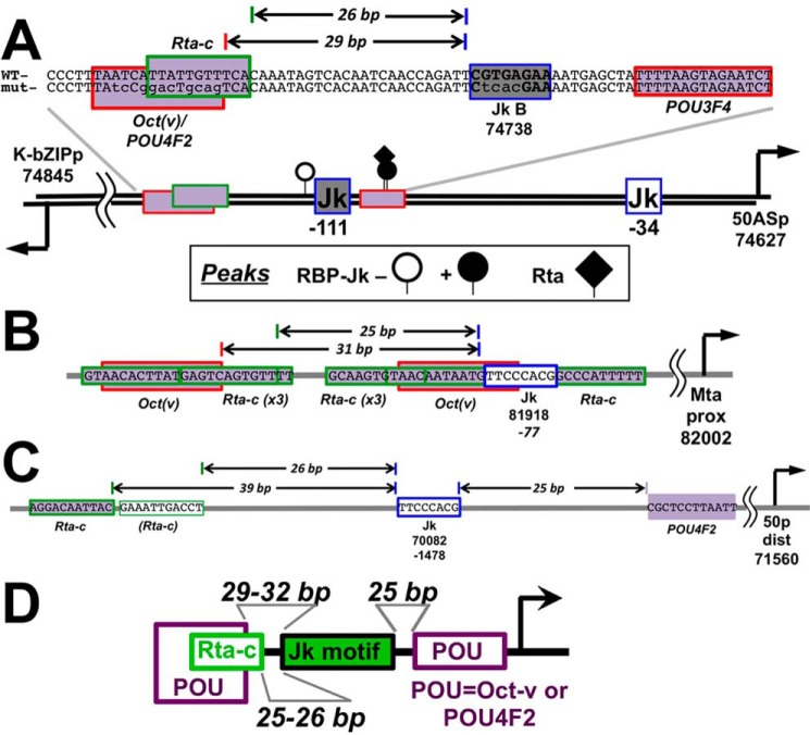
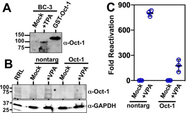
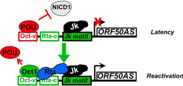
Reactivation of Kaposi's sarcoma-associated herpesvirus (KSHV) from latency requires the viral transactivator Rta to contact the host protein Jκ recombination signal-binding protein (RBP-Jκ or CSL). RBP-Jκ normally binds DNA sequence-specifically to determine the transcriptional targets of the Notch-signaling pathway, yet Notch alone cannot reactivate KSHV. We previously showed that Rta stimulates RBP-Jκ DNA binding to the viral genome. On a model viral promoter, this function requires Rta to bind to multiple copies of an Rta DNA motif (called "CANT" or Rta-c) proximal to an RBP-Jκ motif. Here, high-resolution ChIP/deep sequencing from infected primary effusion lymphoma cells revealed that RBP-Jκ binds nearly exclusively to different sets of viral genome sites during latency and reactivation. RBP-Jκ bound DNA frequently, but not exclusively, proximal to Rta bound to single, but not multiple, Rta-c motifs. To discover additional regulators of RBP-Jκ DNA binding, we used bioinformatics to identify cellular DNA-binding protein motifs adjacent to either latent or reactivation-specific RBP-Jκ-binding sites. Many of these cellular factors, including POU class homeobox (POU) proteins, have known Notch or herpesvirus phenotypes. Among a set of Rta- and RBP-Jκ-bound promoters, Rta transactivated only those that also contained POU motifs in conserved positions. On some promoters, POU factors appeared to inhibit RBP-Jκ DNA binding unless Rta bound to a proximal Rta-c motif. Moreover, POU2F1/Oct-1 expression was induced during KSHV reactivation, and POU2F1 knockdown diminished infectious virus production. Our results suggest that Rta and POU proteins broadly regulate DNA binding of RBP-Jκ during KSHV reactivation.

Keywords: DNA viruses; DNA-binding protein; KSHV; Notch pathway; Oct-1; POU2F1; Rta; chromatin immunoprecipitation (ChiP); herpesvirus; tumor virus; viral DNA.


Conflict of interest statement

The authors declare that they have no conflicts of interest with the contents of this article

Figures

Figure 1.
Figure 1.
DNA elements required for Rta stimulation of RBP-Jκ DNA binding to the Mta promoter. A, promoter schematic and Rta-c (CANT motif) element logo as in Ref. . Jk, RBP-Jκ motif; R, Rta-c (CANT motif); inverted triangle, A/T3 trinucleotide in repeat; arrow, transcription start site. B, sequence logos for RBP-Jκ. Consensus logos as in Ref. and HOCOMOCO as in Refs. , . Y axes indicate bits for all logos.
Figure 2.
Figure 2.
ChIP/Seq of Rta and RBP-Jκ DNA binding on the KSHV genome during latency and reactivation. Visualization of ChIP/Seq peaks by read depth per bp using Integrative Genomics Viewer (IGV) (85). A, genome positions from bp 1 to 68,000. B, genome positions from bp 68,001 to 137,508. Antibodies used are indicated at right, with chromatin from latent (−VPA) or reactivated (+VPA) virus in BC-3 cells. Solid arrows represent open reading frames (ORFs). Numbering above arrows indicate ORF names. Purple arrowheads and two-headed arrows indicate 15 co-localized Rta and RBP-Jκ peaks in reactivation. Asterisks indicate peak positions mapped to PAN promoter (PAN), ORF50AS/K-bZIP promoter (K-bZIP), and Mta promoter (Mta). Orange bars indicate positions of ori-Lyts.
Figure 3.
Figure 3.
qPCR confirmation of selected ChIP/Seq peaks. Cross-linked chromatin was prepared from triplicate samples of untreated and VPA-treated BC-3 cells at 24 h post-VPA addition. A, Rta antibody was used to precipitate chromatin from VPA-treated cells. B, RBP-Jκ antibody was used to precipitate chromatin from untreated (−VPA) and VPA-treated (+VPA) cells. Chromatin precipitated with anti-Rta or anti-RBP-Jκ was quantitated by qPCR using primers corresponding to each of the promoters indicated below the graphs, normalized to qPCR from chromatin precipitated by control rabbit IgG, and then expressed as a proportion of qPCR from input chromatin. Thick lines indicate means of values; thin lines indicate standard errors; + or − above each of the bars indicates whether the region amplified corresponded to the summit of a ChIP/Seq peak in Fig. 1.
Figure 4.
Figure 4.
Fraction of RBP-Jκ peaks mapped to DE or L promoters, co-located with Rta peaks and with Mta promoter motifs during latency and reactivation. Green segments represent RBP-Jκ peaks that increase in reactivation; red segments represent RBP-Jκ peaks that decrease in reactivation, and black segments are peaks that are unchanged in reactivation. Numbers of peaks in each category are indicated. A, proportions of all RBP-Jκ peaks in each category and those in DE or L promoters. B, same as A, but showing only the subset of RBP-Jκ peaks with co-localized Rta peaks. C, same as A, but showing subsets of RBP-Jκ peaks associated with each motif and/or Rta peak, as indicated.
Figure 5.
Figure 5.
Sequence logos for POU DNA motifs. POU motifs from HOCOMOCO (48, 49), aligned to Oct-v motif (50) and Rta-c (CANT) DNA consensus (16) in inverted orientation.
Figure 6.
Figure 6.
Candidate promoter segments selected from ChIP experiments. A, genomic locations. Top shows the name and location of each segment. Bottom shows the visualization of ChIP/Seq peaks using IGV (85). Antibodies used are indicated at right, and chromatin was from latent (−VPA) or reactivated (+VPA) virus in BC-3 cells. Solid arrows represent open reading frames (ORFs). Numbering above arrows indicates ORF name. Peak heights are given in read depth per genomic position. Purple arrowheads below each panel indicate co-localized Rta and RBP-Jκ peaks in reactivation. B, portions of candidate promoters. Schematics representing portions of candidate promoters containing RBP-Jκ motifs and RBP-Jκ peaks are shown. The names of each promoter and the boundaries of schematics are indicated at left. The boxed legend explains the symbols. Arrows indicate positions of transcription start sites mapped to the viral genome within each candidate promoter. Motif text colors are as follows: red = over-represented at RBP-Jκ latency peaks; green = over-represented at RBP-Jκ reactivation peaks; purple = motif over-represented at coincident RBP-Jk/Rta peaks. Activated and nonactivated refer to results of Rta transactivation shown in Fig. 7.
Figure 7.
Figure 7.
RBP-Jκ DNA binding is not sufficient for Rta transactivation. A, ORF50AS promoter. Reporter plasmid was co-electroporated into BL-41 cells alone or with increasing amounts of Rta expression vector in triplicate. Cells were harvested 48 h post-electroporation, and luciferase was measured. Each luciferase value was normalized to β-gal expression from a second reporter plasmid that was co-electroporated as control. Fold transactivation was calculated by comparison with the promoter reporter transfected alone. Inset, typical results of Western blotting to show expression of ectopic Rta in transactivation experiments. Top panel, Western blotting of total cellular protein probed with anti-Rta serum. Bottom panel, Western blotting from same gel probed with anti-actin. B, other candidate promoters. Each of the indicated reporter plasmids were co-electroporated into BL-41 cells with Rta expression vector following the procedure described in A. The maximal transactivation from each titration curve was normalized to transactivation of the Mta proximal (Mta prox) promoter, which was set at 100%. Thick lines indicate means of values, and thin lines indicate standard errors. Chart at top shows result of Rta + Jk binding from Fig. 2, and the presence or absence of indicated motifs as in Fig. 6. dist prox, distal and proximal.
Figure 8.
Figure 8.
Six predicted RBP-Jκ motifs bind to purified RBP-Jκ protein. A, sequences of EMSA oligos chosen from promoters in Fig. 6. Top (+) and bottom (−) strands are indicated. B and C, competition EMSAs. Increasing amounts of the indicated and unlabeled competitor DNAs were preincubated with GST-RBP-Jκ, before addition of 32P-labeled Mta proximal (Mta prox) DNA. Mixtures were electrophoresed and visualized by autoradiography. Vertical line in B indicates cropping of irrelevant lanes in center of image. D, quantitation. Shifted complexes from each gel in B were measured by phosphorimaging. Each complex was normalized to the complexes formed by GST-RBP-Jκ and labeled Mta-proximal DNA alone, in each gel, which was set as 100%. Blue text indicates RBP-Jκ motifs from promoters activated by Rta in Fig. 7. prox dist, proximal distal.
Figure 9.
Figure 9.
Rta-c/Oct-v motif is necessary for optimal transactivation of the ORF50AS/K-bZIP promoter by Rta. WT and mutant reporters were tested for Rta transactivation using the approach described in the legend to Fig. 8, A and B. The sequence of the Rta-c/Oct-v motif in the ORF50AS/K-bZIP promoter is shown at top. Mutated bps are indicated by lowercase lightface type. A, basal activity of promoter/reporters. B, fold transactivation by Rta. Thick lines indicate means of values, and thin lines indicate standard errors.
Figure 10.
Figure 10.
RBP-Jκ element location is important for Rta transactivation of the ORF50AS and K-bZIP promoters. A, transactivation of the WT and mutant ORF50AS and K-bZIP promoters. Each RBP-Jκ–binding site in the promoter was mutated alone, or together, and then tested for Rta transactivation using the approach described in the legend to Fig. 7, A and B. Thick lines indicate means of values, and thin lines indicate standard errors. B, distance of the RBP-Jκ motif to the Rta-c/Oct-v motif determines Rta transactivation magnitude. Each RBP-Jκ–binding site in the promoter was altered as shown in the schematic and then tested for Rta transactivation using the approach described in the legend to Fig. 7, A and B. For both panels, results are shown as percentage of transactivation of WT promoters by Rta divided by empty vector. Promoter schematics follow the design described in the legend to Fig. 6. Thick lines indicate means of values, and thin lines indicate standard errors.
Figure 11.
Figure 11.
Architecture of RBP-Jκ, POU, and Rta-c motifs are similar in the ORF50AS/K-bZIP (A), Mta (B), and ORF50 (C) distal promoters. Established or predicted cis-regulatory elements are shown by rectangles. The arrows on the thick line in A indicate positions of DE transcriptional start sites, with genomic coordinates listed below (8). Numbers below each RBP-Jκ motif (blue outlined Jk) indicate genomic coordinates and/or positions of motifs relative to transcriptional start sites. Horizontal arrows on DNA sequences indicate bp distances between motifs. D, summary of motif arrangements in Rta-transactivated promoters.
Figure 12.
Figure 12.
Oct-1 is required for optimal KSHV reactivation. A, Oct-1 is induced during KSHV reactivation in PEL cells. KSHV-infected BC-3 cells were treated with TPA or with vehicle control (mock) and tested for Oct-1 expression by Western blotting. Purified recombinant GST–Oct-1 protein was included as a Western blotting control. Numbers at left indicate positions of protein apparent molecular weight standards. B, Oct-1 siRNA specifically knocks down Oct-1. Whole-cell protein extracts from Vero cells transfected in Fig. 10A were pooled, and equal concentrations were analyzed by SDS-PAGE/Western blotting. RRL = rabbit reticulocyte lysate programmed to express Oct-1 protein. The asterisk indicates migration of Oct-1–specific protein band. Numbers at left indicate positions of protein apparent molecular weight standards. C,. Oct-1 knockdown debilitates KSHV reactivation. KSHV-infected Vero rKSHV.294 cells were transfected with Oct-1–specific siRNA and control siRNA (scrambled), incubated for 48 h, and then treated with 1.0 mm VPA for 6 h. Media were replaced with fresh media and incubated an additional 48 h. Infectious virus was quantitated by SeAP assay. Fold reactivation was calculated by normalizing SeAP activity to that for control cells, which had been incubated with supernatants from Vero rKSHV.294 cells that were transfected with control siRNA without VPA. Thick lines indicate means of values, and thin lines indicate standard errors. Nontarg = transfected with control (nontargeting) siRNAs.
Figure 13.
Figure 13.
Model for regulation of RBP-Jκ DNA binding on the ORF50AS promoter. POU proteins inhibit NICD1/RBP-Jκ interaction in latency. Rta/Oct-1 complex removed POU and sustains and stimulates Jk DNA binding in reactivation.

Similar articles

Cited by

References

    1. Carbone A., Gaidano G., Gloghini A., Larocca L. M., Capello D., Canzonieri V., Antinori A., Tirelli U., Falini B., and Dalla-Favera R. (1998) Differential expression of BCL-6, CD138/syndecan-1, and Epstein-Barr virus-encoded latent membrane protein-1 identifies distinct histogenetic subsets of acquired immunodeficiency syndrome-related non-Hodgkin's lymphomas. Blood 91, 747–755 - PubMed
    1. Gramolelli S., and Schulz T. F. (2015) The role of Kaposi sarcoma-associated herpesvirus in the pathogenesis of Kaposi sarcoma. J. Pathol. 235, 368–380 10.1002/path.4441 - DOI - PubMed
    1. Mesri E. A., Cesarman E., and Boshoff C. (2010) Kaposi's sarcoma and its associated herpesvirus. Nat. Rev. Cancer 10, 707–719 10.1038/nrc2888 - DOI - PMC - PubMed
    1. Lukac D. M., Kirshner J. R., and Ganem D. (1999) Transcriptional activation by the product of open reading frame 50 of Kaposi's sarcoma-associated herpesvirus is required for lytic viral reactivation in B cells. J. Virol. 73, 9348–9361 - PMC - PubMed
    1. Lukac D. M., Renne R., Kirshner J. R., and Ganem D. (1998) Reactivation of Kaposi's sarcoma-associated herpesvirus infection from latency by expression of the ORF 50 transactivator, a homolog of the EBV R protein. Virology 252, 304–312 10.1006/viro.1998.9486 - DOI - PubMed

Publication types

MeSH terms

Substances