

Neural substrates of sexual arousal are not sex dependent
- PMID: 31308220
- PMCID: PMC6681749
- DOI: 10.1073/pnas.1904975116
Neural substrates of sexual arousal are not sex dependent
Abstract
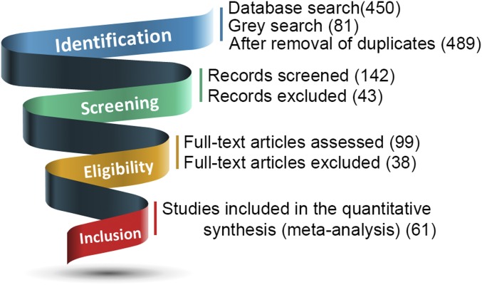
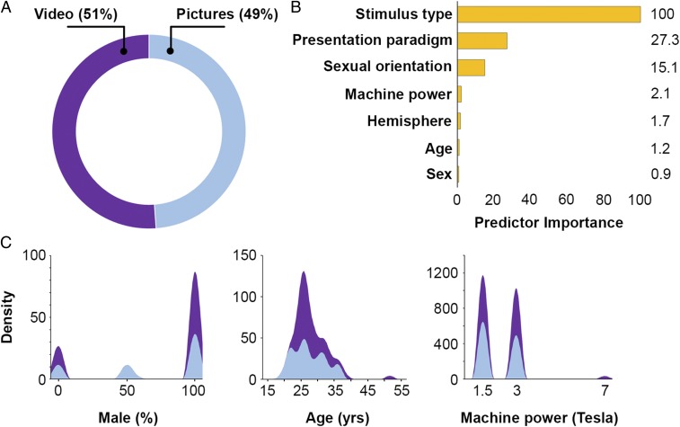
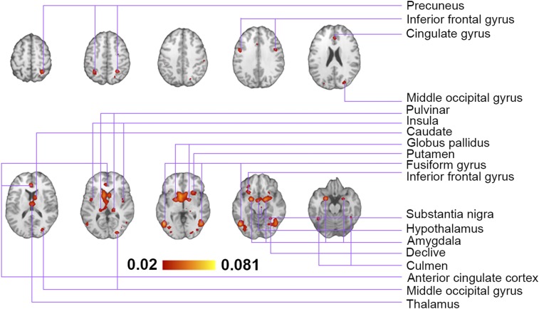
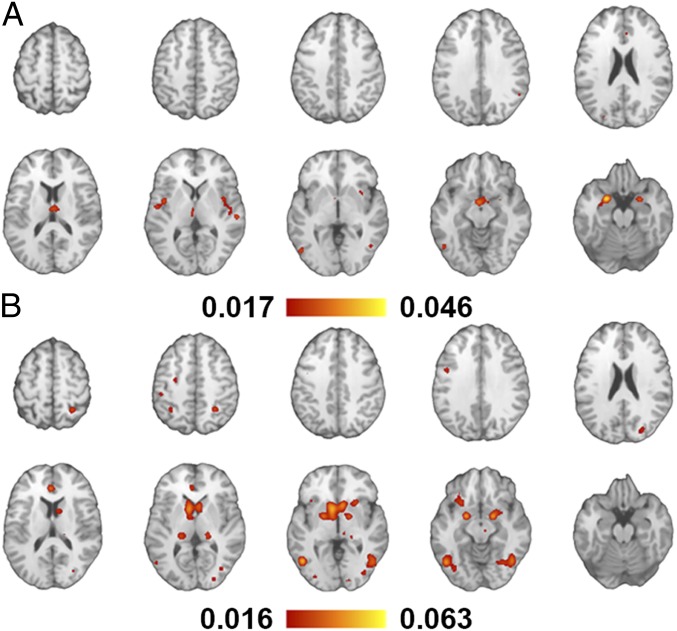
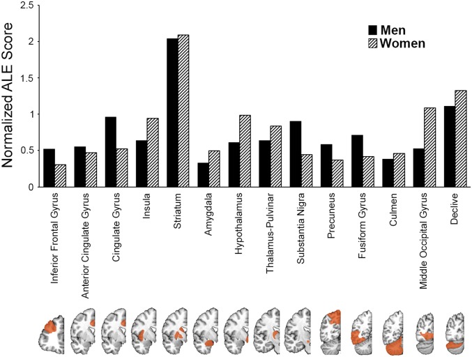
Sexual arousal is a dynamical, highly coordinated neurophysiological process that is often induced by visual stimuli. Numerous studies have proposed that the cognitive processing stage of responding to sexual stimuli is the first stage, in which sex differences occur, and the divergence between men and women has been attributed to differences in the concerted activity of neural networks. The present comprehensive metaanalysis challenges this hypothesis and provides robust quantitative evidence that the neuronal circuitries activated by visual sexual stimuli are independent of biological sex. Sixty-one functional magnetic resonance imaging studies (1,850 individuals) that presented erotic visual stimuli to men and women of different sexual orientation were identified. Coordinate-based activation likelihood estimation was used to conduct metaanalyses. Sensitivity and clustering analyses of averaged neuronal response patterns were performed to investigate robustness of the findings. In contrast to neutral stimuli, sexual pictures and videos induce significant activations in brain regions, including insula, middle occipital, anterior cingulate and fusiform gyrus, amygdala, striatum, pulvinar, and substantia nigra. Cluster analysis suggests stimulus type as the most, and biological sex as the least, predictor for classification. Contrast analysis further shows no significant sex-specific differences within groups. Systematic review of sex differences in gray matter volume of brain regions associated with sexual arousal (3,723 adults) did not show any causal relationship between structural features and functional response to visual sexual stimuli. The neural basis of sexual arousal in humans is associated with sexual orientation yet, contrary to the widely accepted view, is not different between women and men.
Keywords: metaanalysis; neuroimaging; sex differences; sexual arousal.
Copyright © 2019 the Author(s). Published by PNAS.
Conflict of interest statement
The authors declare no conflict of interest.
Figures
Comment in
-
Reply to Poeppl et al.: Controlling for false positive rates is critical for accurate and consistent interpretation of findings.Proc Natl Acad Sci U S A. 2020 May 26;117(21):11206. doi: 10.1073/pnas.1916336117. Epub 2020 May 5. Proc Natl Acad Sci U S A. 2020. PMID: 32371492 Free PMC article. No abstract available.
-
Neural substrates of sexual arousal revisited: Dependent on sex.Proc Natl Acad Sci U S A. 2020 May 26;117(21):11204-11205. doi: 10.1073/pnas.1915927117. Epub 2020 May 5. Proc Natl Acad Sci U S A. 2020. PMID: 32371493 Free PMC article. No abstract available.
References
-
- Wellings K., et al. , Sexual behaviour in context: A global perspective. Lancet 368, 1706–1728 (2006). - PubMed
-
- Chung W. S., Lim S. M., Yoo J. H., Yoon H., Gender difference in brain activation to audio-visual sexual stimulation; do women and men experience the same level of arousal in response to the same video clip? Int. J. Impot. Res. 25, 138–142 (2013). - PubMed
-
- Schmidt G., Male-female differences in sexual arousal and behavior during and after exposure to sexually explicit stimuli. Arch. Sex. Behav. 4, 353–365 (1975). - PubMed
-
- Steinman D. L., Wincze J. P., Sakheim D. K., Barlow D. H., Mavissakalian M., A comparison of male and female patterns of sexual arousal. Arch. Sex. Behav. 10, 529–547 (1981). - PubMed
-
- Laan E., Everaerd W., van Bellen G., Hanewald G., Women’s sexual and emotional responses to male- and female-produced erotica. Arch. Sex. Behav. 23, 153–169 (1994). - PubMed
Publication types
MeSH terms
Substances
LinkOut - more resources
Full Text Sources
Medical
