. 2019 Nov;18(11):2310-2323.
doi: 10.1074/mcp.TIR119.001433. Epub 2019 Jul 15.

A Platform for Extracellular Interactome Discovery Identifies Novel Functional Binding Partners for the Immune Receptors B7-H3/CD276 and PVR/CD155

Affiliations

A Platform for Extracellular Interactome Discovery Identifies Novel Functional Binding Partners for the Immune Receptors B7-H3/CD276 and PVR/CD155

Bushra Husain et al. Mol Cell Proteomics. 2019 Nov.

Abstract

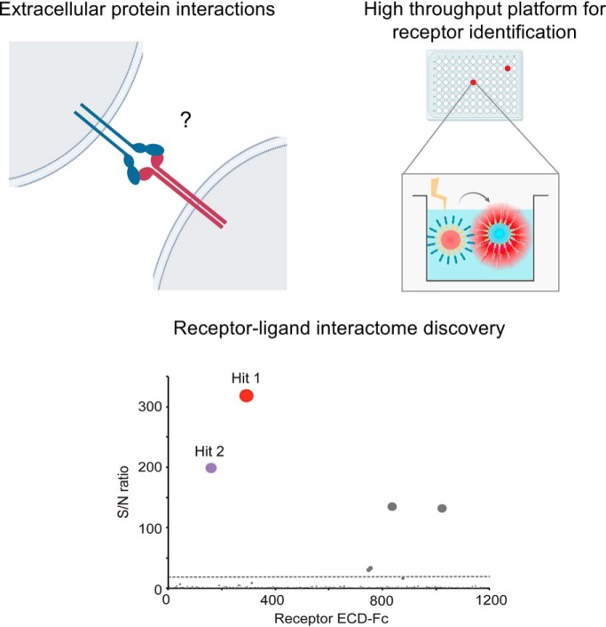
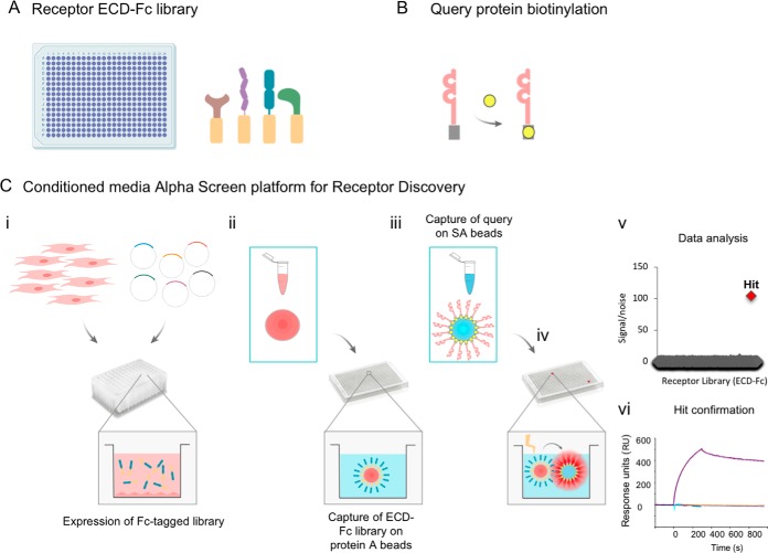
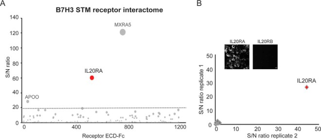
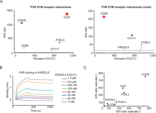
Receptors expressed on the plasma membrane and their interacting partners critically regulate cellular communication during homeostasis and disease, and as such represent main therapeutic targets. Despite its importance for drug development, receptor-ligand proteomics has remained a daunting field, in part because of the challenges associated to the study of membrane-expressed proteins. Here, to enable sensitive detection of receptor-ligand interactions in high throughput, we implement a new platform, the Conditioned Media AlphaScreen, for interrogation of a library consisting of most single transmembrane human proteins. Using this method to study key immune receptors, we identify and further validate the interleukin receptor IL20RA as the first binding partner for the checkpoint inhibitor B7-H3. Further, KIR2DL5, a natural killer cell protein that had remained orphan, is uncovered as a functional binding partner for the poliovirus receptor (PVR). This interaction is characterized using orthogonal assays, which demonstrate that PVR specifically engages KIR2DL5 on natural killer cells leading to inhibition of cytotoxicity. Altogether, these results reveal unappreciated links between protein families that may importantly influence receptor-driven functions during disease. Applicable to any target of interest, this technology represents a versatile and powerful approach for elucidation of receptor-ligand interactomes, which is essential to understand basic aspects of the biology of the plasma membrane proteins and ultimately inform the development of novel therapeutic strategies.

Keywords: assay development; automation; cancer therapeutics; cell-cell interactions; protein-protein interactions; secretome.


Figures

None
Graphical abstract
Fig. 1.
Fig. 1.
Conditioned Media AlphaScreens for sensitive detection of transient receptor-ligand interactions. Biotinylated PD-1 protein was incubated with different amounts of streptavidin-coated donor beads (16 to 0 μg) at the molar concentrations indicated (50–0 nm). Binding to the known interacting partners A, PD-L1 or B, PD-L2 was analyzed using the procedure indicated Experimental procedures. PD-L1, PD-L2, or a control protein (labeled as “C”) expressed as recombinant ECD fused to a Fc tag, were captured on protein A-coated acceptor beads by incubating with different amounts of beads (16 to 0 μg). For these assays, PD-L1 and PD-L2 were tested at 10 nm concentration. Based on these results, 4 μg of beads were used for subsequent assays to minimize reagent consumption while allowing detection of low affinity PPIs with high signal/noise ratios. Plots show one representative assay out of two independent runs, assayed in triplicates. C–E, Biotinylated PD-1 (50 nm concentration) was captured on donor beads to analyze binding to acceptor beads that had been incubated with of conditioned media enriched in C, PD-L1, D, PD-L2 or E, a control receptor, expressed as ECD-Fc fusions. Fc-tagged receptors ECD in the conditioned media were titrated and diluted to the indicated concentrations (0–5 μg/ml) to assess sensitivity of the binding assays. Experiments shown are representative of at least two independent assays run in triplicates. Error bars indicate S.D.
Fig. 2.
Fig. 2.
Automated Conditioned Media AlphaScreens workflow. A, A library consisting of ≈1,200 unique single transmembrane (STM) human receptors, expressed as extracellular domains fused to a Fc tag, was generated. B, The query protein under study is expressed as a recombinant protein for in vitro biotinylation, or expressed as an avi-tagged protein for site-specific biotinylation. C, Schematic representation of the Conditioned Media AlphaScreen platform for receptor interactome discovery: i, The STM receptor library is expressed using a high throughput platform for small-scale cell transfection. ii, Conditioned media enriched in individual receptor-Fc proteins is harvested and transferred to multi-well plates, following incubation with protein A-coated acceptor beads. iii, The biotinylated query protein is captured on streptavidin-coated donor beads and iv, incubated with the library of STM receptors, previously immobilized on acceptor beads. v, Query protein-receptor interactions are detected by reading the chemiluminescence signal, followed by data analysis for identification of interacting partners for the query protein under study. vi, Confirmation of binding partners of interest using additional methodologies such as biolayer interferometry.
Fig. 3.
Fig. 3.
Conditioned Media AlphaScreen for high sensitivity elucidation of immune receptor interactomes. Recombinant PD-1 and CD86 proteins were biotinylated and analyzed for binding to the library of STM receptors following the experimental procedure described in the text. S/N ratio ≥ 20 was determined as experimentally-validated cut-off for hit calling (dotted gray line). A, PD-1 screens using the Conditioned Media AlphaScreen platform identify PD-L1 and PD-L2 and B, CD86 screening using the Conditioned Media AlphaScreen workflow allows identification of the binding partners CTLA4 and CD28 as top scoring hits. Hits labeled in gray color have been empirically determined to be nonspecific binders.
Fig. 4.
Fig. 4.
The interleukin receptor IL20RA is a receptor for the immune checkpoint B7-H3. A, B7-H3 was expressed as a recombinant protein and screened using the Conditioned Media AlphaScreen technology. The B7-H3 STM receptor interactome identifies IL20RA as a top hit. Proteins labeled in gray color represent nonspecific binders. S/N ratio ≥ 20 was used as experimentally-determined cut-off for hit calling (dotted gray line). B, B7-H3 binding to IL20RA on the cell surface. Binding of recombinant biotinylated B7-H3 to cells expressing IL20RA or control receptors (IL20RA, IL20RB, IL17RB, IL10RA, IL10RB, LIFR, OSMR, and IL6R) was analyzed by immunofluorescence, using fluorophore-conjugated streptavidin for detection. Intersection plot shows B7-H3 binding to the surface of cells expressing the indicated receptors; transfections were performed in duplicates. Micrographs show images corresponding to B7-H3 binding to IL20RA- and IL20RB-expressing cells, as control. Scale bar = 50 μm.
Fig. 5.
Fig. 5.
The orphan NK cell receptor KIR2DL5A is a novel binding partner for PVR. A, PVR screens using the Conditioned Media AlphaScreens platform identify known interacting partners as well as a previously unknown interaction with the receptor KIR2DL5A (inset). B, Analysis of PVR binding to KIR2DL5A, expressed as recombinant purified proteins, by BLI. Biotinylated PVR was immobilized on streptavidin sensors and tested for binding to KIR2DL5, as a soluble ECD-Fc tag analyte, at the indicated concentrations. C, PVR binding to receptors expressed on the cell surface. Individual KIR receptors (KIR2DL1, KIR2DL2, KIR2DL3, KIR2DL4, KIR2DL5A, KIR2DL5B, KIR2DS1, KIR2DS3, KIR2DS4, KIR2DS5, KIR3DL1, KIR3DL3) or known PVR binders (CD96, CD226, TIGIT, PVRL3, and PVRL4) were transiently expressed in cells, and binding of PVR to the cell surface was analyzed by immunofluorescence using fluorescent streptavidin for detection. Binding is represented as signal intensity/noise, and shown as intersection plots representing two independent transfections. Representative images for PVR binding to the cell surface are shown.
Fig. 6.
Fig. 6.
PVR modulates NK cell cytotoxicity in a KIR2DL5A-dependent manner. A, Sorted KIR2DL5+ (blue) or KIR2DL5 (red) LAK cells were used as effector cells against A-427 targets at a ratio of 2:1. Data are shown as % cytotoxicity determined from cell index values measured every 10 min over a 4 h period. Each time point is mean ± S.D. of duplicate wells. Real-time profiling of cytotoxicity shown is representative of three independent experiments. B, Sorted KIR2DL5+ (blue) or KIR2DL5 (red) LAK cells were pre-incubated with anti-KIR2DL5 (clone UP-R1) antibody before addition to A-427 target cells. Data shown is representative of two independent experiments. C, Sorted KIR2DL5+ (blue) or KIR2DL5 (red) LAK cells were tested against A-427(ΔPVR/ΔPVRL2) target cells. Data shown is representative of two independent experiments. D, Comparison of LAK-mediated killing at 4 h. Compiled data for KIR2DL5+ (blue) or KIR2DL5 (red) LAK cell killing against A-427 WT target cells (solid bars, n = 3), A-427 WT cytotoxicity in the presence of anti-KIR2DL5 antibody (hatched bars, n = 2), or killing of A-427.DKO targets (open bars, n = 2). E, Effect of CD226 antibody blockade. KIR2DL5+ (blue) or KIR2DL5 (red) LAK cell killing against A-427 WT in the absence (solid bars) or presence (dotted bars) of anti-CD226 antibody at 4 h. Data shown is representative of two independent experiments. F, Blocking of KIR2DL5A binding to PVR-expressing cells in the presence of an anti-KIR2DL5A antibody. KIR2DL5A binding to the cell surface is represented as mean fluorescence intensity (MFI) relative to binding in the absence of antibody. Data represents mean ± S.D. of duplicate wells. G, PVR binding to CD226-expressing cells in the presence of KIR2DL5A, TIGIT or CD226, expressed as recombinant proteins. Biotinylated PVR was incubated alone or in the presence of the PVR binders at the indicated molar ratios, and PVR bound to the surface was detected with fluorophore-conjugated streptavidin. Data shown is representative of two independent experiments.
Fig. 7.
Fig. 7.
KIR2DL5 modulates NK cell effector function upon interaction with PVR on the cell surface. A, Recognition of PVR on the tumor cell by the activating receptor CD226 on the NK cell results in cell activation and killing. B, NK cell-expressed KIR2DL5 binds to PVR, even in the presence of CD226 co-expressed on the tumor cell, delivering an inhibitory signal that reduces NK-mediated cytolysis of the tumor cell. C, Antibody blockade of KIR2DL5 restores cytotoxic functions and killing of the tumor cell.

Similar articles

Cited by

References

    1. Hopkins A. L., and Groom C. R. (2002) The druggable genome. Nat. Rev. Drug Discov. 1, 727–730 - PubMed
    1. Overington J. P., Al-Lazikani B., and Hopkins A. L. (2006) How many drug targets are there? Nat. Rev. Drug Discov. 5, 993–996 - PubMed
    1. Wright G. J., Martin S., Bushell K. M., and Sollner C. (2010) High-throughput identification of transient extracellular protein interactions. Biochem. Soc. Trans. 38, 919–922 - PubMed
    1. Martinez-Martin N. (2017) Technologies for proteome-wide discovery of extracellular host-pathogen interactions. J. Immunol. Res. 2017, 2197615. - PMC - PubMed
    1. Nooren I. M., and Thornton J. M. (2003) Diversity of protein-protein interactions. EMBO J. 22, 3486–3492 - PMC - PubMed