

Anti-Jo-1 Antibody-positive Interstitial Pneumonia in an Elderly Patient with Congenital Rubella Syndrome
- PMID: 31308343
- PMCID: PMC6701999
- DOI: 10.2169/internalmedicine.2454-18
Anti-Jo-1 Antibody-positive Interstitial Pneumonia in an Elderly Patient with Congenital Rubella Syndrome
Abstract
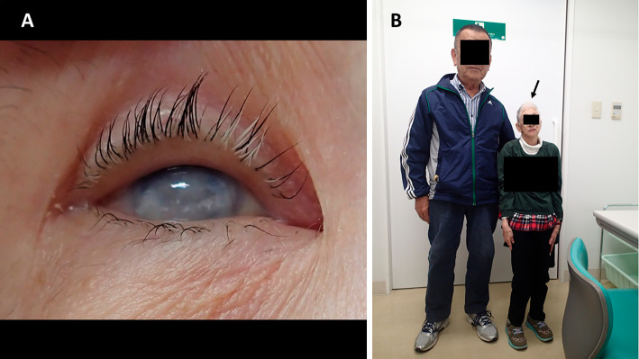
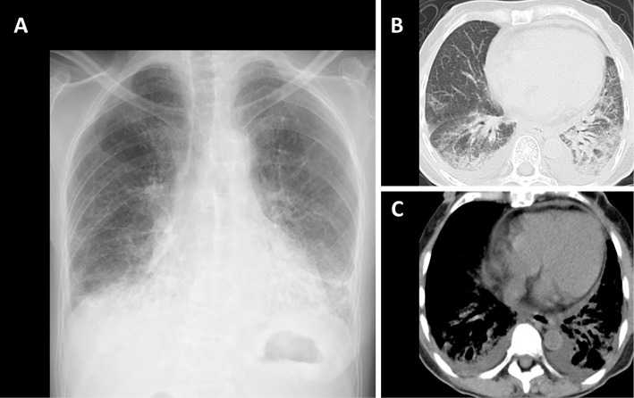
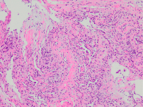
A 71-year-old woman with congenital rubella syndrome (CRS) presented with prolonged cough. No physical findings suggested the presence of any connective tissue diseases. Chest computed tomography showed ground-glass opacities and consolidations in the bilateral lower lobes. She had elevated serum Krebs von den Lungen-6, hypoxemia and positive serum anti-Jo-1 antibody. Bronchoalveolar lavage fluid revealed lymphocytosis with a decreased CD4/CD8 ratio. A transbronchial lung biopsy specimen revealed organizing pneumonia. Based on a diagnosis of interstitial pneumonia with autoimmune features (IPAF), systemic corticosteroids were administered, and a good outcome was obtained. A possible relationship between CRS and IPAF is herein discussed.
Keywords: autoimmunity; congenital rubella syndrome; interstitial pneumonia.
Conflict of interest statement
Figures
Similar articles
-
Bucillamine-induced Pneumonitis in a Patient with Rheumatoid Arthritis-associated Interstitial Pneumonia: A Case Report and Review of the Literature.Intern Med. 2019 Aug 1;58(15):2207-2211. doi: 10.2169/internalmedicine.2515-18. Epub 2019 Mar 28. Intern Med. 2019. PMID: 30918198 Free PMC article.
-
Serological and morphological prognostic factors in patients with interstitial pneumonia with autoimmune features.BMC Pulm Med. 2017 Aug 14;17(1):111. doi: 10.1186/s12890-017-0453-z. BMC Pulm Med. 2017. PMID: 28807021 Free PMC article.
-
Patterns of interstitial lung disease during everolimus treatment in patients with metastatic renal cell carcinoma.Jpn J Clin Oncol. 2012 May;42(5):442-6. doi: 10.1093/jjco/hys033. Epub 2012 Mar 20. Jpn J Clin Oncol. 2012. PMID: 22438405
-
[Interstitial lung disease in connective tissue diseases].Rev Prat. 2007 Dec 31;57(20):2235-42. Rev Prat. 2007. PMID: 18320743 Review. French.
-
Interstitial Pneumonia with Autoimmune Features and Undifferentiated Connective Tissue Disease.Semin Respir Crit Care Med. 2019 Apr;40(2):271-277. doi: 10.1055/s-0039-1684007. Epub 2019 May 28. Semin Respir Crit Care Med. 2019. PMID: 31137065 Review.
Cited by
-
Prevalence and Characteristics of Rheumatoid-Associated Autoantibodies in Patients with COVID-19.J Inflamm Res. 2021 Jul 12;14:3123-3128. doi: 10.2147/JIR.S312090. eCollection 2021. J Inflamm Res. 2021. PMID: 34285538 Free PMC article.
References
-
- Sugishita Y, Shimatani N, Katow S, Takahashi T, Hori N. Epidemiological characteristics of rubella and congenital rubella syndrome in the 2012-2013 epidemics in Tokyo, Japan. Jpn J Infect Dis 68: 159-165, 2015. - PubMed
-
- Sugishita Y, Akiba T, Sumitomo M, et al. . Shedding of rubella virus among infants with congenital rubella syndrome born in Tokyo, Japan, 2013-2014. Jpn J Infect Dis 69: 418-423, 2016. - PubMed
-
- Boner A, Wilmott RW, Dinwiddie R, et al. . Desquamative interstitial pneumonia and antigen-antibody complexes in two infants with congenital rubella. Pediatrics 72: 835-839, 1983. - PubMed
-
- Franklin SL, Kelley R. Congenital rubella and interstitial pneumonitis. Clin Pediatr (Phila) 40: 101-103, 2001. - PubMed
-
- Sanchez MO, Chang AB. Congenital rubella pneumonitis complicated by Pneumocystis jiroveci infection with positive long term respiratory outcome: a case report and literature review. Pediatr Pulmonol 44: 1235-1239, 2009. - PubMed
Publication types
MeSH terms
Substances
LinkOut - more resources
Full Text Sources
Medical
Research Materials