. 2019 Jul 15;10(8):539.
doi: 10.1038/s41419-019-1774-z.

The Kindlin2-p53-SerpinB2 signaling axis is required for cellular senescence in breast cancer

Affiliations

The Kindlin2-p53-SerpinB2 signaling axis is required for cellular senescence in breast cancer

Khalid Sossey-Alaoui et al. Cell Death Dis. .

Abstract

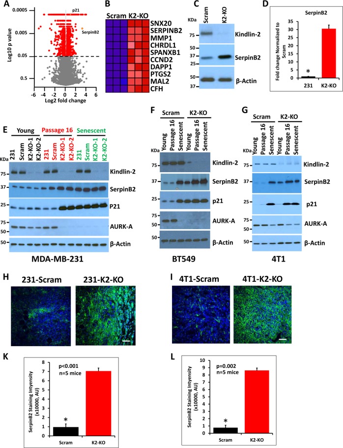
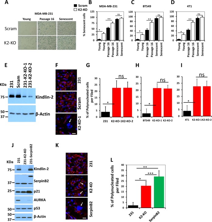
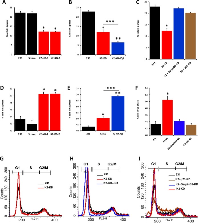
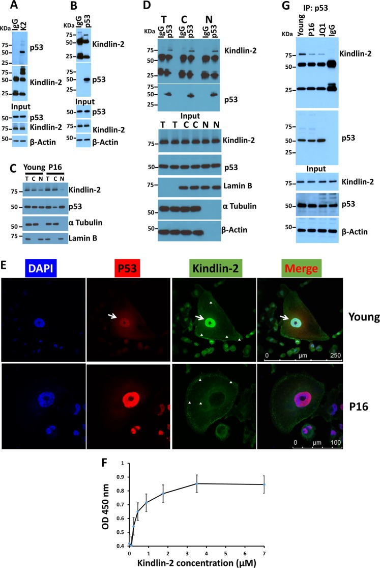
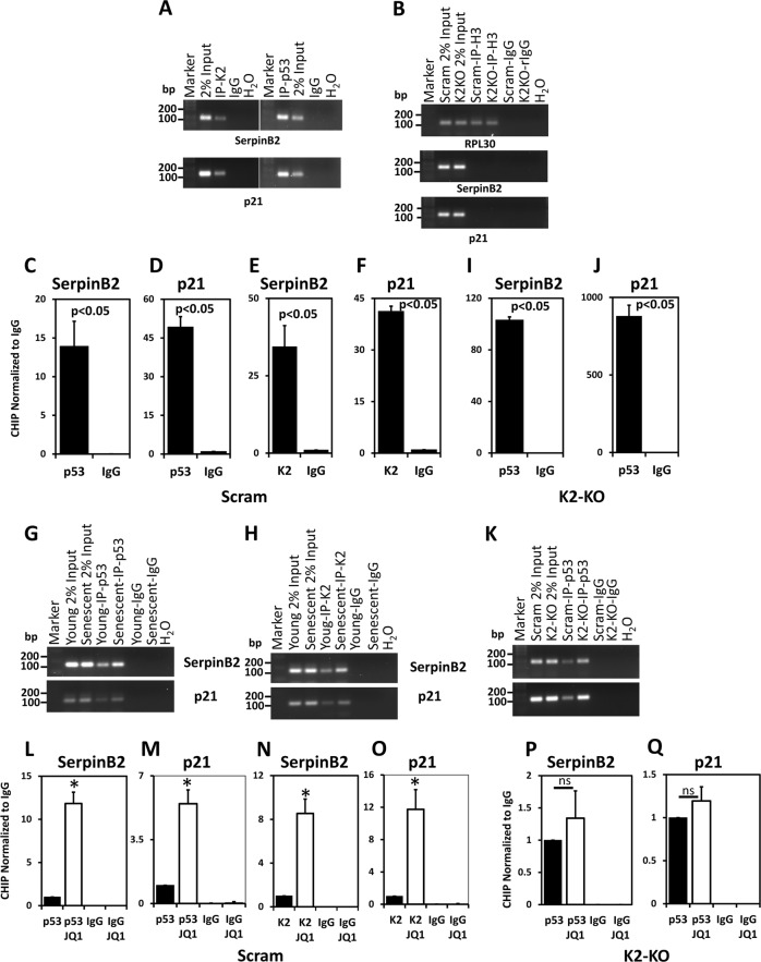
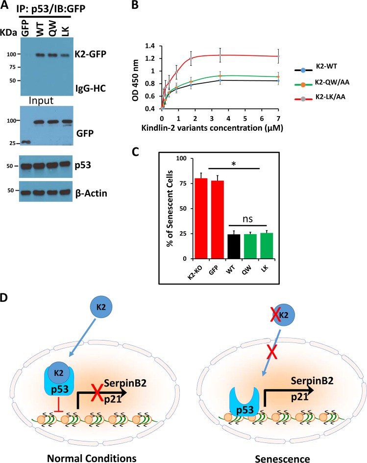
In cancer, cellular senescence is a complex process that leads to inhibition of proliferation of cells that may develop a neoplastic phenotype. A plethora of signaling pathways, when dysregulated, have been shown to elicit a senescence response. Two well-known tumor suppressor pathways, controlled by the p53 and retinoblastoma proteins, have been implicated in maintaining the cellular senescence phenotype. Kindlin-2, a member of an actin cytoskeleton organizing and integrin activator proteins, has been shown to play a key role in the regulation of several hallmarks of several cancers, including breast cancer (BC). The molecular mechanisms whereby Kindlin-2 regulates cellular senescence in BC tumors remains largely unknown. Here we show that Kindlin-2 regulates cellular senescence in part through its interaction with p53, whereby it regulates the expression of the p53-responsive genes; i.e., SerpinB2 and p21, during the induction of senescence. Our data show that knockout of Kindlin-2 via CRISPR/Cas9 in several BC cell lines significantly increases expression levels of both SerpinB2 and p21 resulting in the activation of hallmarks of cellular senescence. Mechanistically, interaction between Kindlin-2 and p53 at the promotor level is critical for the regulated expression of SerpinB2 and p21. These findings identify a previously unknown Kindlin-2/p53/SerpinB2 signaling axis that regulates cellular senescence and intervention in this axis may serve as a new therapeutic window for BCs treatment.


Conflict of interest statement

The authors declare that they have no conflict of interest.

Figures

Fig. 1
Fig. 1. Loss of Kindlin-2 induces breast cancer cell senescence by activating SerpinB2 in vitro and in vivo.
a Volcano plot from the RNA-seq analysis of the differentially expressed genes between control (Scram) and pooled cell populations of Kindlin-2-deficient (KO-KO) MDA-MB-231 cells. The X-axis represents the Log2 fold change in expression levels and the Y-axis shows the p values. p21 and SerpinB2 are shown in orange dots. b Heat-map showing the top 10 differentially expressed genes between control (Scram) and Kindlin-2-deficient (K2-KO) MDA-MB-231 cells (Passage 16). c Western blots with anti-kindlin-2 (top panel) and anti-SerpinB2 (middle panel) on lysates of control (Scram) and Kindlin-2-deficient (K2-KO) MDA-MB-231 cells. Anti-β-Actin (lower panel) was used as a loading control. d Quantitative real-time RT-PCR of SerpinB2 transcripts in control (Scram) and Kindlin-2-deficient (K2-KO) MDA-MB-231 cells. Data are representative of 3 independent experiments (ns, not significant); *, **p < 0.05, student t-test. e-g Western blots with the indicated antibodies on lysates of parental (231), control (Scram) and Kindlin-2-deficient (K2-KO) MDA-MB-231 e, BT549 f or 4T1 cells g at passage 3 or less (Young), passage 16 or treated with 1 mM JQ1 for 10 days (Senescent). Anti-β-Actin was used as a loading control. h, i Representative images of control (Scram) or Kindlin-2-deficient (K2-KO) MDA-MB-231 h or 4T1 i -derived tumor sections stained for SerpinB2 (green). Nuclei were counterstained with DAPI. Scale bar, 146 μm. k, l Quantification of SerpinB2 staining areas in MDA-MB-231 k and 4T1 l. Data are expressed as mean ± SEM. *p < 0.005; n = 5 mice
Fig. 2
Fig. 2. Senescence-associated β-galactosidase activity is activated in Kindlin-2-deficient breast cancer cells, and loss of Kindlin-2 induces polynucleation of BC cells that can be enhanced by overexpression of SerpinB2.
a Representative micrographs of young, passage 16 and senescent control (scram) or Kindlin-2-deficient (K2-KO) MDA-MB-231 cells that were stained for senescence-associated (SA) β-galactosidase activity (blue staining). b–d Quantification of the % of cells of SA-β-galactosidase activity in MDA-MB-231 b, BT549 c, and 4T1 cells d. Data are representative of 3 independent experiments (ns, not significant; *, **p < 0.05, student t-test). e Western blots with the indicated antibodies on lysates of parental (231), control (Scram), Kindlin-2-deficient (K2-KO1 and K2-KO-2) cells. Anti-β-Actin (lower panel) was used as a loading control. f Representative confocal microscopy micrographs of parental (231, top panel), control (Scram, middle panel) or Kindlin-2-deficient (K2-KO-1, lower panel) MDA-MB-231 cells that were stained for actin filament (red). Nuclei were counterstained with DAPI (blue). White arrows point to polynucleated cells. Scale bar, 20 μm. g–i Quantification of the % of polynucleated cells in MDA-MB-231 g, BT549 h, and 4T1 i cells. j Western blots with the indicated antibodies on lysates of parental (231), Kindlin-2-deficient (K2-KO) or SerpinB2-overexpressing MDA-MB-231 (231-SepinB2) cells. Anti-β-Actin (lower panel) was used as a loading control. k Representative confocal microscopy micrographs of parental (231, top panel), Kindlin-2-deficient (K2-KO, middle panel) or SerpinB2-overexpressing MDA-MB-231 (SepinB2, bottom panel) cells that were stained for actin filament (red). Nuclei were counterstained with DAPI (blue). White arrows point to polynucleated cells. Scale bar, 20 μm. l Quantification of the % of polynucleated cells shown in Fig. 3g. Data are representative of 3 independent experiments (*, **, ***p < 0.05, student t-test)
Fig. 3
Fig. 3. Kindlin-2 is required for the orderly progression of the cell cycle.
a Quantification of percent of cells in S phase from parental (231), control shRNA(Scram) or Kindlin-2-knockdown MDA-MB-231 cells using K2-shRNA clone 1 (K2-KD-1) or K2-shRNA clone 2 (K2-KD-2). b Quantification of percent of cells in S phase from parental (231), Kindlin-2-knockdown MDA-MB-231 cells using K2-shRNA clone 1 (K2-KD-1), or K2-KD MDA-MB-231 cells treated with 1 mM JQ1. c Quantification of percent of cells in S phase from parental (231), Kindlin-2-knockdown MDA-MB-231 cells using K2-shRNA clone 1 (K2-KD-1), SerpinB2-Knockdown (SerpinB2-KD) or p21-knockdown (p21-KD). SerpinB2-Knockdown and p21-knockdown were performed in K2-deficient MDA-MB-231 (231-K2-KD). d Quantification of percent of cells in G1 phase from parental (231), control (Scram) or Kindlin-2-knockdown MDA-MB-231 cells using K2-shRNA clone 1 (K2-KD-1) or K2-shRNA clone 2 (K2-KD-2). e Quantification of percent of cells in G1 phase from parental (231), Kindlin-2-knockdown MDA-MB-231 cells using K2-shRNA clone 1 (K2-KD-1), or K2-KD MDA-MB-231 cells treated with 1 mM JQ1. f Quantification of percent of cells in G1 phase from parental (231), Kindlin-2-knockdown MDA-MB-231 cells using K2-shRNA clone 1 (K2-KD-1), SerpinB2-Knockdown (SerpinB2-KD) or p21-knockdown (p21-KD). Data are representative of 3 independent experiments (*, **, ***p < 0.05, student t-test). g–i Representative histograms using flow cytometry of parental (231), control shRNA (Scram) or the indicated gene knockdowns and treatments
Fig. 4
Fig. 4. Kindlin-2 interacts with p53 in both the cytoplasm and the nucleus of cancer cells.
a, b Protein lysates prepared from early passage (young) MDA-MB-231 cells was used for immunoprecipitation with mouse anti-Kindlin-2 antibody or control mouse IgG a; and mouse anti-p53 antibody or control mouse IgG b and subjected to immunoblotting analysis with rabbit anti-p53 antibody (a, upper panel) or mouse anti-Kindlin-2 antibody (b, upper panel). In control blots, cell lysates were also immunoblotted with mouse anti-Kindlin-2 and rabbit anti-p53 antibodies to show the presence of equal amounts of these proteins in the cell lysates (input panels). β-Actin is a loading control. c Total, nuclear and cytoplasmic fractions of protein lysates from early passage (young) and senescent (P16) MDA-MB-231 cells were subjected to immunoblotting with the indicated antibodies. d Total, nuclear and cytoplasmic fractions of protein lysates from early passage (young) MDA-MB-231 cells were used for immunoprecipitation with mouse anti-p53 antibody or control mouse IgG, and subjected to immunoblotting analysis with mouse anti-Kindlin-2 antibody (upper panel) or rabbit anti-p53 antibody (upper panel). In control blots (input panels), protein lysates fractions were also immunoblotted with mouse anti-Kindlin-2 and rabbit anti-p53 antibodies to show the presence of equal amounts of these proteins in the lysates as well as the specificity for the nuclear (Lamin B) and the cytoplasmic (α Tubulin). β-Actin is a loading control. e Confocal microscopy images of immunofluorescence staining of early passage (young) or senescent (P16) MDA-MB-231 cells that were stained for p53 (Red) or Kindlin-2 (Green). Nuclei were counterstained with DAPI. White arrows point to the co-localization of Kindlin-2 and p53 in the nucleus of young but not senescent cells in the merged image. The white arrowheads point to the localization of Kindlin-2 to focal adhesions in both young and senescent cells. Higher magnifications of other representative images are shown in Supplemental Fig. 2. f Interaction of Kindlin-2-GST with purified p53. The binding isotherms of increasing concentrations of GST-tagged WT Kindlin-2 to the wells of microtiter plates coated with p53. The data are expressed as means ± SEM of triplicates of two representative experiments. g Protein lysates prepared from early passage (young), passage 16 (P16) or JQ-1 treated (1 mM for 2 days) MDA-MB-231 cells was used for immunoprecipitation with mouse anti-p53 antibody or control mouse IgG and subjected to immunoblotting analysis with rabbit anti-Kindlin-2 antibody or rabbit anti-p53 antibody (upper panels). In control blots (input panels), cell lysates were also immunoblotted with mouse anti-Kindlin-2 and rabbit anti-p53 antibodies to show the presence of equal amounts of these proteins in the cell lysates. β-Actin is a loading control
Fig. 5
Fig. 5. Kindlin-2 binds to p53 to regulate expression of SerpinB2 and p21 at the promoter level.
a Semi-quantitative agarose gel PCR of CHIP of MDA-MD-231 cells using either anti-Kindlin-2 (left panels) or anti-p53 (right panels) followed by PCR amplification of SepinB2 promoter (upper panel) or p21 promoter (lower panels). The corresponding IgG was used as a negative control for the immunoprecipitation and 2% input of the DNA-chromatin complex was used as a positive control for the PCR. b Control CHIP and PCR experiment using anti-Histone H3 for immunoprecipitation of chromatin-DNA complex from control (Scram) or Kindlin-2-deficient (K2-KO) MDA-MB-231 cells, followed by Semi-quantitative agarose gel PCR for the promoter of RPL30 (upper panel), SerpinB2 (middle panel) or p21 (lower panel). Only the RPL30 promoter was amplified in the H3-immunoprecipitates. IgG was used as a negative control for the immunoprecipitation and 2% input of the DNA-chromatin complex was used as a positive control for the PCR. c–f Quantitative real-time PCR of DAN-chromatin immunoprecipitates of control (Scram) MDA-MB-231 cells using anti-p53 c, f or anti-Kindlin-2 e, f, followed by PCR of SepinB2 c, e and p21 d. f promoter, and normalized to the CHIP PCR of the corresponding IgG. i, j Quantitative real-time PCR of DNA-chromatin immunoprecipitates of Kindlin-2-deficient (K2-KO) MDA-MB-231 cells using anti-p53, followed by PCR of SepinB2 promoter i and p21 promoter j, and normalized to the CHIP PCR of the corresponding IgG. g–k Semi-quantitative agarose gel PCR CHIP of young or senescent MDA-MD-231 cells using either anti-p53 g or anti-Kindlin-2 h, or Kindlin-2-deficient MDA-MB-231 cells using anti-p53 k, followed by PCR amplification of SepinB2 promoter (upper panels) or p21 promoter (lower panels). The corresponding IgG was used as a negative control for the immunoprecipitation and 2% input of the DNA-chromatin complex was used as a positive control for the PCR. l–o Quantitative real-time PCR of DNA-chromatin immunoprecipitates of untreated or JQ1-treated control (Scram) MDA-MB-231 cells using anti-p53 l, m or anti-Kindlin-2 n, o, followed by PCR of SepinB2 promoter l, n and p21 promoter m, o, and normalized to the CHIP PCR of the corresponding IgG. (p and q) Quantitative real-time PCR of DNA-chromatin immunoprecipitates of untreated or JQ1-treated Kindlin-2-deficient (K2-KO) MDA-MB-231 cells using anti-p53, followed by PCR of SepinB2 promoter p and p21 promoter q, and normalized to the CHIP PCR of the corresponding IgG. Data are representative of 3 independent experiments (*p < 0.05, student t-test)
Fig. 6
Fig. 6. The Kindlin-2-mediated regulation of senescence may not depend on its binding to integrin or actin
a Kindlin-2 deficient (K2-KO) MDA-MB-231cells were transiently transfected with expression vectors for control GFP, wild-type Kindlin-2, QW/AA-Kindlin-2 or LK/AA-Kindlin-2 mutants fused to GFP. The corresponding total protein lysates were used for immunoprecipitation with rabbit anti-p53 antibody, and subjected to immunoblotting analysis with mouse anti-GFP antibody (upper panel). In control blots (Input blots), the same cell lysates were also immunoblotted with mouse anti-GFP and rabbit anti-p53 antibodies to show the presence of equal amounts of these proteins in the cell lysates. β-Actin is a loading control. b Interaction of Kindlin-2-GST with purified p53. The binding isotherms of increasing concentrations of GST-tagged WT Kindlin-2 and its mutant variants Kindlin-2 (QW/AA) and Kindlin-2 (LK/AA) to the wells of microtiter plates coated with p53. The data are expressed as means ± SEM of triplicates of two representative experiments. c Quantification of SA β-galactosidase staining in the Kindlin-2-deficient (K2-KO) MDA-MB-23 and the K2-KO cells transfected with the control GFP, wild type Keindlin-2 (WT), QW/AA (QW) or LK/AA (LK) Kindlin-2 mutants. Data are representative of 3 independent experiments (*p < 0.05, student t-test). d Model describing the role of Kindlin-2 in the regulation of senescence in breast cancer

References

    1. Hayflick L, Moorhead PS. The serial cultivation of human diploid cell strains. Exp. Cell Res. 1961;25:585–621. doi: 10.1016/0014-4827(61)90192-6. - DOI - PubMed
    1. Campisi J. Aging, cellular senescence, and cancer. Annu. Rev. Physiol. 2013;75:685–705. doi: 10.1146/annurev-physiol-030212-183653. - DOI - PMC - PubMed
    1. Salama R, Sadaie M, Hoare M, Narita M. Cellular senescence and its effector programs. Genes Dev. 2014;28:99–114. doi: 10.1101/gad.235184.113. - DOI - PMC - PubMed
    1. He S, Sharpless NE. Senescence in health and disease. Cell. 2017;169:1000–1011. doi: 10.1016/j.cell.2017.05.015. - DOI - PMC - PubMed
    1. Vaziri H, Benchimol S. Alternative pathways for the extension of cellular life span: inactivation of p53/pRb and expression of telomerase. Oncogene. 1999;18:7676–7680. doi: 10.1038/sj.onc.1203016. - DOI - PubMed

Publication types

MeSH terms