. 2019 Jul 15;9(1):10241.
doi: 10.1038/s41598-019-46572-3.

Early evaluation of corneal collagen crosslinking in ex-vivo human corneas using two-photon imaging

Affiliations

Early evaluation of corneal collagen crosslinking in ex-vivo human corneas using two-photon imaging

Ana Batista et al. Sci Rep. .

Abstract

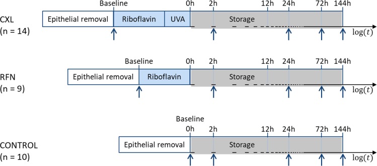
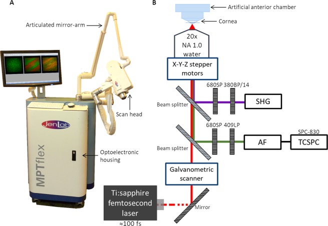
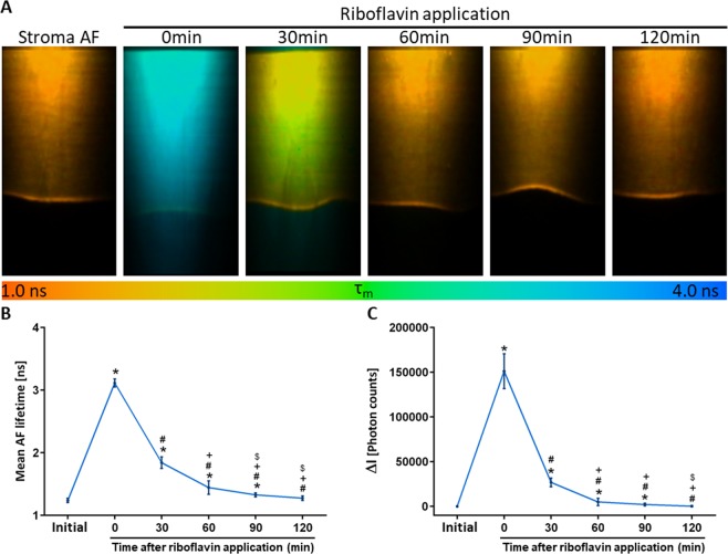
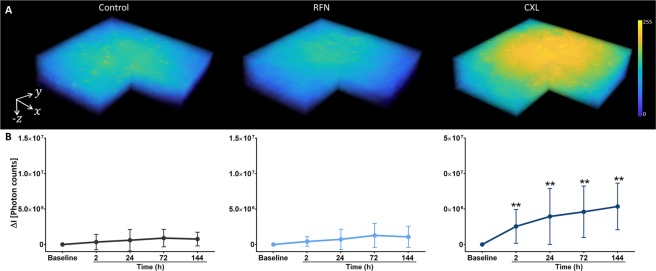
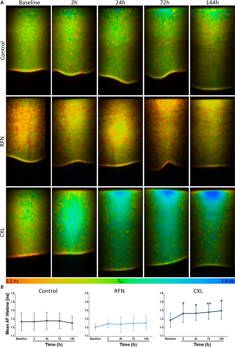
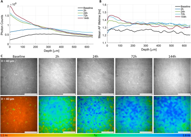
The clinical outcome of corneal collagen crosslinking (CXL) is typically evaluated several weeks after treatment. An earlier assessment of its outcome could lead to an optimization of the treatment, including an immediate re-intervention in case of failure, thereby, avoiding additional discomfort and pain to the patient. In this study, we propose two-photon imaging (TPI) as an earlier evaluation method. CXL was performed in human corneas by application of riboflavin followed by UVA irradiation. Autofluorescence (AF) intensity and lifetime images were acquired using a commercial clinically certified multiphoton tomograph prior to CXL and after 2h, 24h, 72h, and 144h storage in culture medium. The first monitoring point was determined as the minimum time required for riboflavin clearance from the cornea. As control, untreated samples and samples treated only with riboflavin (without UVA irradiation) were monitored at the same time points. Significant increases in the stroma AF intensity and lifetime were observed as soon as 2h after treatment. A depth-dependent TPI analysis showed higher AF lifetimes anteriorly corresponding to areas were CXL was most effective. No alterations were observed in the control groups. Using TPI, the outcome of CXL can be assessed non-invasively and label-free much sooner than with conventional clinical devices.


Conflict of interest statement

The authors declare no competing interests.

Figures

Figure 1
Figure 1
Schematic representation of the timing of crosslinking experiments. The vertical arrows indicate time points of two-photon imaging. CXL – corneal collagen crosslinking, RFN – riboflavin.
Figure 2
Figure 2
Multiphoton tomograph MPTflex (JenLab GmbH, Berlin, Germany) used for image acquisition (A) and schematic representation of the instrumental optical setup (B).
Figure 3
Figure 3
Cross-sectional autofluorescence (AF) lifetime images of the cornea during riboflavin clearance (A). Average changes of the mean AF lifetime (B) and of the AF intensity variation (ΔI) over time (C). Cross-sections cover an area of 600×1200 μm2. Statistical significance was computed using Mann Whitney U test. *statistically significant at ρ < 0.05 compared with baseline; #statistically significant at ρ < 0.05 compared with t = 0 min; +statistically significant at ρ < 0.05 compared with t = 30 min; $statistically significant at ρ < 0.05 compared with t = 60 min.
Figure 4
Figure 4
3D representations of the donor corneal samples for control, RFN and CXL groups after 72 h storage. Volumes cover 300 × 300 × 300 μm3 and were reconstructed from individual and sequential autofluorescence (AF) intensity images recorded with 5 μm intervals along the z axis. Average differences on AF intensities variation (ΔI) for all groups over time. Statistical significance was computed using t-test. **statistically significant at ρ < 0.01.
Figure 5
Figure 5
Cross-sectional autofluorescence (AF) lifetime images of the cornea at baseline, 2 h, 24 h, 72 h, and 144 h for control, RFN and CXL groups (A) and average variations in the mean AF lifetime over time (B). Cross-sections represent an area of 600 × 1200 μm2. Statistical significance was obtained using t-test. *statistically significant at ρ < 0.05; **statistically significant at ρ < 0.01.
Figure 6
Figure 6
Corneal stroma autofluorescence (AF) intensity (A) and mean AF lifetime (B) after CXL as a function of depth for baseline, 2 h, 24 h, 72 h, and 144 h. AF intensity and corresponding AF lifetime images for the same corneal depth as function of time (C). Scale bar = 100 μm.

References

    1. Hovakimyan M, Guthoff RF, Stachs O. Collagen cross-linking: current status and future directions. J. Ophthalmol. 2012;2012(406850):1–12. doi: 10.1155/2012/406850. - DOI - PMC - PubMed
    1. Zhang X, et al. A review of collagen cross-linking in cornea and sclera. J. Ophthalmol. 2015;2015(289467):1–12. - PMC - PubMed
    1. Zyablitskaya M, et al. Evaluation of therapeutic tissue crosslinking (TXL) for myopia using second harmonic generation signal microscopy in rabbit sclera. Investig. Opthalmology Vis. Sci. 2017;58:21–29. doi: 10.1167/iovs.16-20241. - DOI - PMC - PubMed
    1. Khan YA, et al. Riboflavin and ultraviolet light a therapy as an adjuvant treatment for medically refractive Acanthamoeba keratitis: report of 3 cases. Ophthalmology. 2011;118:324–31. doi: 10.1016/j.ophtha.2010.06.041. - DOI - PubMed
    1. Wollensak G, Spoerl E, Seiler T. Riboflavin/ultraviolet-a–induced collagen crosslinking for the treatment of keratoconus. Am J Ophthalmol. 2003;135:620–627. doi: 10.1016/S0002-9394(02)02220-1. - DOI - PubMed

Publication types