. 2019 Sep;38(36):6323-6337.
doi: 10.1038/s41388-019-0881-8. Epub 2019 Jul 15.

Rgnef promotes ovarian tumor progression and confers protection from oxidative stress

Affiliations

Rgnef promotes ovarian tumor progression and confers protection from oxidative stress

Elizabeth G Kleinschmidt et al. Oncogene. 2019 Sep.

Abstract

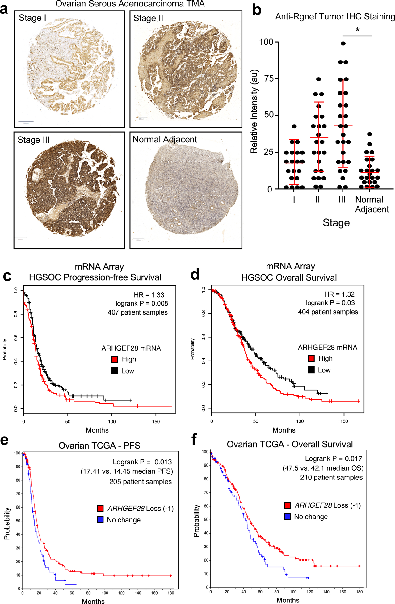
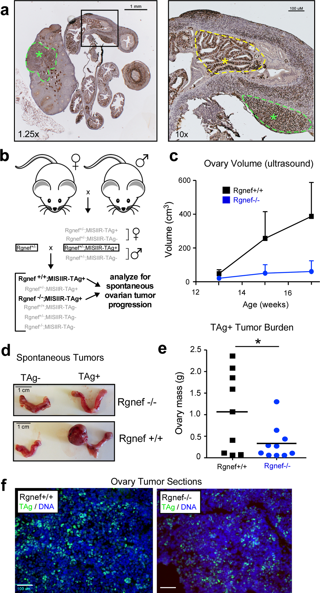
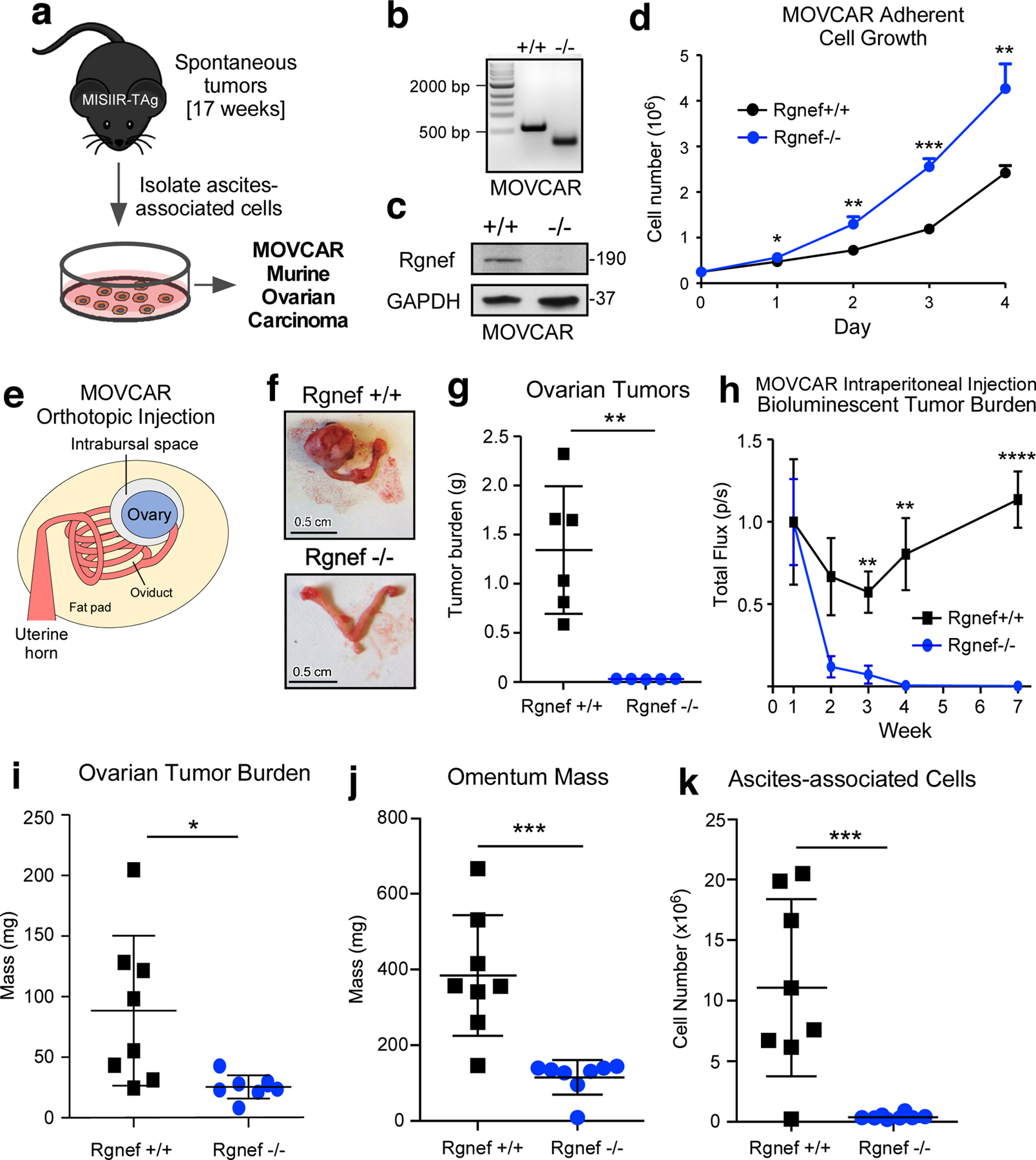
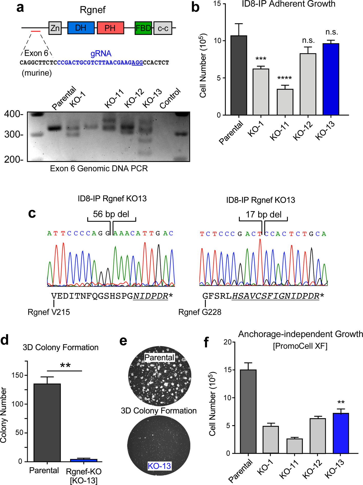
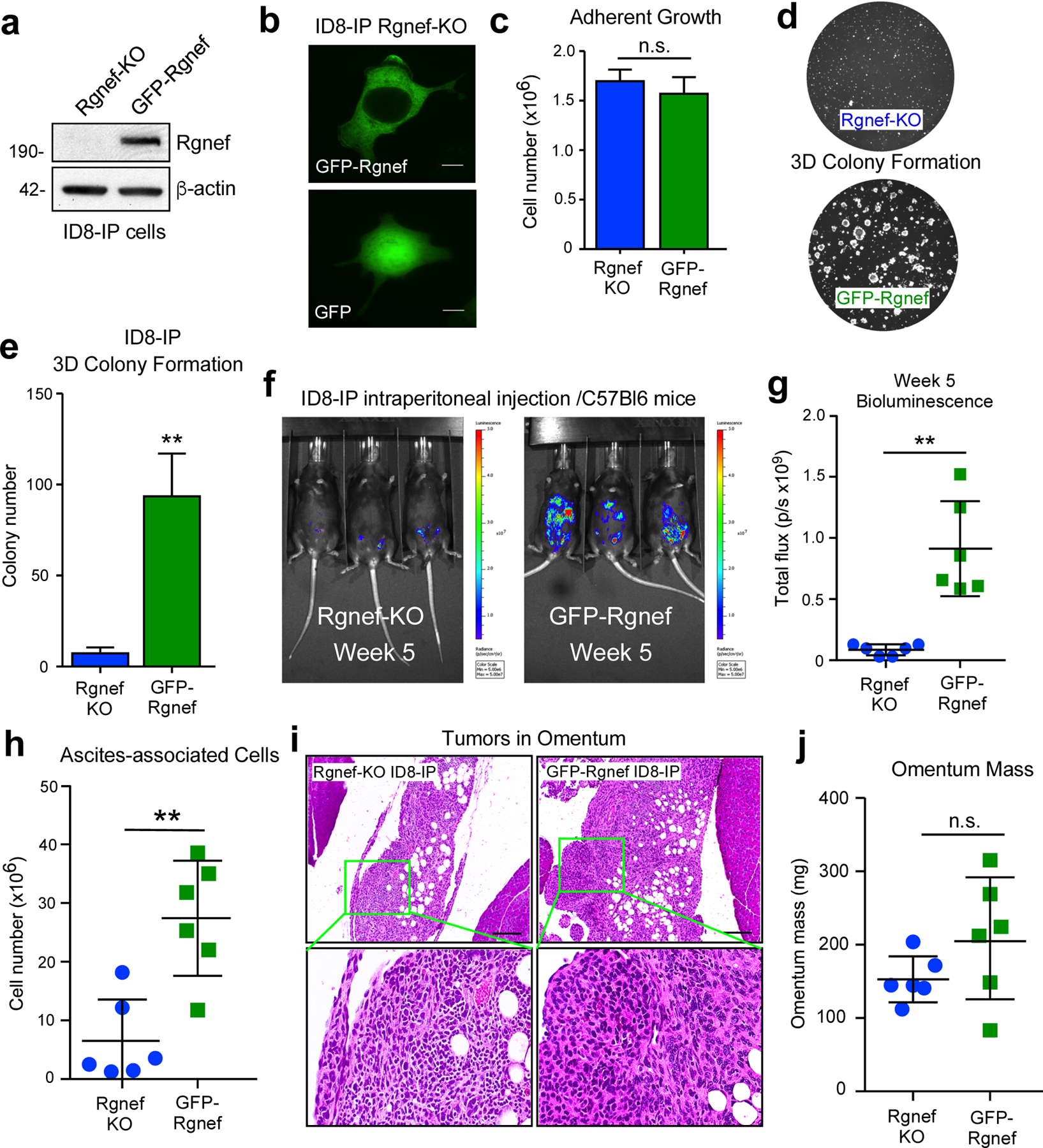
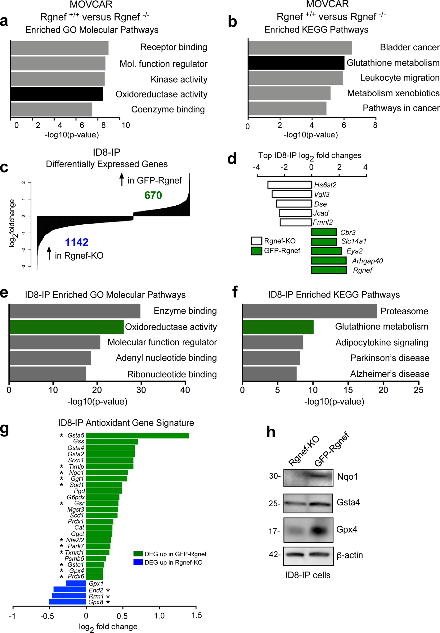
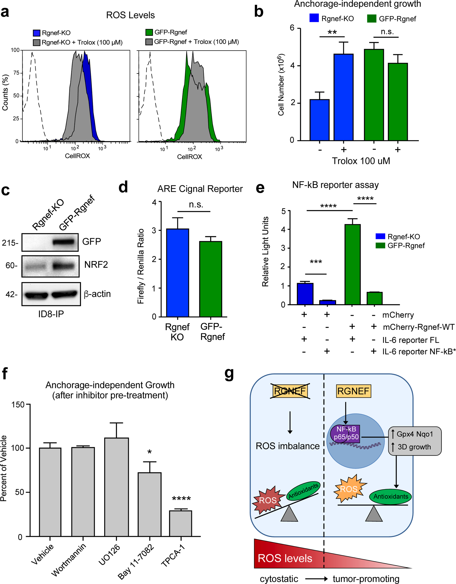
Ovarian cancer is the fifth-leading cause of cancer death among women. The dissemination of ovarian tumors and growth as spheroids accompanies late-stage disease. In cell culture, ovarian tumor cell spheroids can exhibit elevated resistance to environmental stressors, such as reactive oxygen species. Homeostatic balance of the antioxidant response is a protective mechanism that prevents anoikis, a form of programmed cell death. Signaling pathways activated by integrin receptors suppress anoikis. Rgnef (ARHGEF28/p190RhoGEF) is a guanine nucleotide exchange factor that is activated downstream of integrins. We find that Rgnef protein levels are elevated in late-stage serous ovarian cancer, high Rgnef mRNA levels are associated with decreased progression-free and overall survival, and genomic ARHGEF28 loss is associated with increased patient survival. Using transgenic and transplantable Rgnef knockout mouse models, we find that Rgnef is essential for supporting three-dimensional ovarian spheroid formation in vitro and tumor growth in mice. Using RNA-sequencing and bioinformatic analyses, we identify a conserved Rgnef-supported anti-oxidant gene signature including Gpx4, Nqo1, and Gsta4; common targets of the NF-kB transcription factor. Antioxidant treatment enhanced growth of Rgnef-knockout spheroids and Rgnef re-expression facilitated NF-κB-dependent tumorsphere survival. These studies reveal a new role for Rgnef in ovarian cancer to facilitate NF-κB-mediated gene expression protecting cells from oxidative stress.


Conflict of interest statement

Conflicts of Interest: All authors declare no conflicts of interest.

Figures

Figure 1:
Figure 1:
Analysis of Rgnef or ARHGEF28 levels in serous ovarian cancer. (a) Representative high grade serous ovarian cancer tumor micro-array (TMA) cores stained with polyclonal anti-Rgnef antibodies (brown) and nuclei counter-stained with hematoxylin. Stage information was from the manufacturer and normal adjacent is ovarian tissue from tumor-bearing patients. (b) Aperio Image Analysis quantification using the positive pixel count algorithm and relative intensity values (arbitrary units, au) are shown per TMA core. Values are means +/− SD (n=92, P< 0.05, T-Test). (c and d) Kaplan-Meier plotter (http://kmplot.com/ovar) was used to evaluate ARHGEF28 mRNA levels in stage III-IV serous ovarian cancer samples. High ARHGEF28 expression [auto-select cutoff] was significantly associated with (c) progression-free survival (*P = 0.008, n = 407) or (d) overall survival (*P = 0.03, n = 404). Loss of ARHGEF28 at the genomic level (heterozygous loss or homologous deletion vs. maintenance or gain) was similarly evaluated for (e) progression free or overall (f) survival using data available in The Cancer Genome Atlas (TCGA) (*P < 0.02).
Figure 2:
Figure 2:
Rgnef knockout prevents spontaneous ovarian tumor formation. (a) Image of a TAg-stained (brown) ovary and oviduct from a MISIIR-TAg+ mouse. TAg is expressed in the murine ovary and fallopian tubal lesions (green asterisks and dotted lines) and oviduct (yellow asterisk and dotted line). Scale is 1 mm (left) or 100 μM (right). (b) Breeding schematic for generation of Rgnef−/− or Rgnef+/+; MISIIR;TAg+ mice. (c) Ovary size of littermates measured by ultrasound. Mice were sacrificed at week 17 or at earlier humane endpoint. Each point represents n ≥ 5 mice with error bars as SEM. (d) Representative pictures of oviducts and ovaries for each group at endpoint. Scale is 1 cm. (e) Mean tumor burden (total ovary mass) was higher in Rgnef+/+ than Rgnef−/− mice (*P < 0.05, SD +/− 0.93 for Rgnef+/+ and 0.39 for Rgnef−/− mice). (f) Immunofluorescent staining for TAg (green) and DNA (Hoechst, blue) in frozen section of ovaries from Rgnef−/− or Rgnef+/+; MISIIR;TAg+ tumor-bearing mice. Scale is 100 μm.
Figure 3:
Figure 3:
Cell-intrinsic role for Rgnef in promoting murine ovarian cancer (MOVCAR) tumor growth. (a) Schematic of Rgnef+/+ and Rgnef−/− MOVCAR generation. (b) Representative Rgnef exon 24 PCR of MOVCAR cells from Rgnef+/+ and Rgnef−/− TAg+ mice confirming genomic wildtype Rgnef (582 bp) or deletion in Rgnef murine exon 24 (125 bp). (c) Immunoblotting showing Rgnef loss in Rgnef−/− MOVCARs using GAPDH as loading control. (d) Rgnef−/− MOVCARs grow faster in adherent conditions over 4 days (* P ≤ 0.05, ** P ≤ 0.01, ***P ≤0.001, +/−SD, n=3 independent experiments). (e) Schematic of MOVCAR cell orthotopic intrabursal injection into syngeneic MISIIR-TAg-Low; Rgnef+/+ mice. (f) Representative images of oviducts and ovaries from Rgnef+/+ and Rgnef−/− MOVCAR tumor-bearing mice. Scale is 0.5 cm. (g) Quantitation of Rgnef+/+ and Rgnef−/− MOVCAR orthotopic tumor growth (**P ≤ 0.01, +/− SD). (h) Bioluminescent imaging from mice injected intraperitoneally with Rgnef+/+ or Rgnef−/− MOVCARs. Total flux (photons/second) levels are normalized by group to week 1 for each cohort (**P ≤ 0.01, ****P ≤ 0.0001, n=8 for each group, +/− SEM). (i-k) Following intraperitoneal injection, ascites-associated cells were collected at week 14. Rgnef loss impaired metastatic potential as measured by (i) ovarian tumor burden, (j) omentum mass and (k) number of ascites-associated cells (*P ≤ 0.05, ***P ≤ 0.001, +/− SD, n=8 per group).
Figure 4:
Figure 4:
Rgnef loss specifically impairs 3D cell growth. (a) Schematic of CRISPR/Cas9 gRNA targeting of murine Rgnef exon 6 (blue text) within the N-terminal domain. Also shown are Rgnef protein domains: Zn - zinc finger, DH – Dbl homologous, PH - pleckstrin homology, FBD – FAK binding domain, c-c - coiled-coil region. Below: Exon 6 PCR was performed with genomic DNA to identify insertions or deletions in the indicated Rgnef KO clones. Parental ID8-IP cells yield a 393 bp band. (b) Adherent growth of ID8-IP and the indicated Rgnef-KO clones over 4 days (n.s.= no significance, *** P ≤0.001, **** P ≤ 0.0001, n=3 technical replicates, +/− SD). (c) Sanger sequencing of ID8-IP Rgnef KO-13 clone exon 6 identifying two independent deletions (56 and 17 bp) resulting in predicted translational termination. Alternative reading frame is underlined and italicized with position of stop codon (*) denoted (Uniprot protein G5E8P2). (d) ID8-IP and Rgnef KO-13 colony formation (** P ≤ 0.01, n=2 independent experiments, +/− SD). (e) Representative images of colonies in Matrigel. (f) Anchorage-independent growth of ID8-IP and the indicated Rgnef KO clones in serum-free conditions over 5 days (+/− SD, **P ≤ 0.01).
Figure 5:
Figure 5:
Rgnef promotes anchorage-independent growth in vitro and in vivo. (a) Anti-Rgnef immunoblotting showing GFP-Rgnef re-expression in ID8-IP Rgnef-KO cells. Actin is a control. (b) GFP-Rgnef or GFP distribution in ID8-IP Rgnef-KO cells visualized by confocal microscopy. Scale is 2 μm. (c) ID8-IP Rgnef-KO and GFP-Rgnef re-expressing cells exhibit no adherent growth difference (n=3 technical replicates, n.s.= no significance, +/− SD). (d) Colony growth is enhanced by GFP-Rgnef re-expression. Representative images (d) and quantification (e) are shown (**P ≤ 0.01, n=3 biological replicates, +/− SD). (f) Representative bioluminescent imaging of ID8-IP Rgnef-KO or GFP-Rgnef re-expressing cells after 5 weeks. (g) Bioluminescent flux quantified on experimental Day 34 (**P ≤ 0.01, n = 6, +/− SD). (h) Quantitation of ascites-associated cells recovered at Day 39 (**P ≤ 0.01, n = 6, +/− SD). (i) Representative H&E stained images of ID8-IP Rgnef-KO or GFP-Rgnef tumor implants within the omentum. Scale is 50 μm. Inset, high magnification. (j) Final omentum mass from ID8-IP Rgnef-KO or GFP-Rgnef tumor-bearing mice. Differences are not significant (n.s.)
Figure 6:
Figure 6:
Rgnef promotes an antioxidant gene signature. (a) Differentially upregulated mRNAs in MOVCAR Rgnef+/+ as compared to Rgnef−/− were determined using Illumina BeadChip Array and processed by Gene Set Enrichment Analysis (GSEA). Top 5 most enriched GO molecular function (a) or KEGG pathways (b) in the set of 313 upregulated transcripts are shown. (c) RNA sequencing was used to determine differential mRNA levels in Rgnef-KO and GFP-Rgnef re-expressing ID8-IP cells. 670 targets were upregulated in the GFP-Rgnef re-expressing, and 1142 targets were upregulated in Rgnef-KO cells. (d) The top log2 fold changes in the ID8-IP Rgnef-KO or GFP-Rgnef re-expressing cells are shown. (e) Top 5 most enriched GO molecular function or (f) KEGG pathways in the set of 670 upregulated genes in the ID8-IP GFP-Rgnef re-expressing cells. Enrichment score was determined using GSEA. (g) Differentially expressed genes were compared to a curated list of antioxidant genes (Supplemental Table 2). Genes differentially upregulated (green) or downregulated (blue) in ID8-IP GFP-Rgnef cells vs Rgnef-KO cells are shown. (h) Immunoblotting shows increased levels of Nqo1, Gsta4, and Gpx4 in lysates of anchorage-independent (5 days) ID8-IP GFP-Rgnef cells with β-actin as a loading control.
Figure 7:
Figure 7:
Rgnef supports NF-κB activation needed for ID8-IP anchorage-independent cell growth. (a) Cellular reactive oxygen species (ROS) levels in ID8-IP Rgnef-KO and GFP-Rgnef re-expressing cells as measured by CellROX staining with or without antioxidant Trolox (100 μM, 1 h) addition. Flow cytometry histograms are shown. Negative control (dotted line). (b) Trolox increases ID8-IP Rgnef-KO growth in suspension (** P ≤ 0.01, n=3 independent experiments, +/− SD). (c) Immunoblot of ID8-IP Rgnef KO and GFP-Rgnef cell lysates for GFP, NRF2, and actin protein levels. (d) GFP-Rgnef expression did not enhance ARE (antioxidant response element) reporter for NRF2 transcriptional activity in human 293T cells (n.s.= no significance, +/− SD, n=3 technical replicates). (e) mCherry-Rgnef transfection activates an IL-6 luciferase reporter in human 293T cells dependent on NF-κB DNA binding site integrity (NF-κB*). Dual-luciferase activity was measured and normalized (***P ≤ 0.001, ****P ≤ 0.0001, +/− SD, n=3 technical replicates). (f) Adherent ID8-IP GFP-Rgnef cells were pre-treated with DMSO (vehicle), 1 μM wortmannin, 10 μM U0126, 10 μM Bay 11–7082, or 10 μM TPCA-1 for 24h, washed, and then evaluated for anchorage-independent growth (n.s.=no significance, *P ≤ 0.05, ****P ≤ 0.0001, +/− SD, n=3 technical replicates). (g) Model of Rgnef in mediating cell response to oxidative stress. Upon Rgnef loss, the cell is not able to balance increased ROS resulting from growth in suspension, resulting in oxidative stress. When Rgnef is re-expressed, NF-κB-mediated transcription downstream of Rgnef promotes the expression of an antioxidant gene signature, resulting in redox homeostasis.

References

    1. Siegel RL, Miller KD, Jemal A. Cancer statistics, 2018. CA Cancer J Clin 2018; 68: 7–30. - PubMed
    1. Lengyel E. Ovarian cancer development and metastasis. Am J Pathol 2010; 177: 1053–1064. - PMC - PubMed
    1. Shield K, Ackland ML, Ahmed N, Rice GE. Multicellular spheroids in ovarian cancer metastases: Biology and pathology. Gynecol Oncol 2009; 113: 143–148. - PubMed
    1. Kenny HA, Dogan S, Zillhardt M, A KM, Yamada SD, Krausz T et al. Organotypic models of metastasis: A three-dimensional culture mimicking the human peritoneum and omentum for the study of the early steps of ovarian cancer metastasis. Cancer Treat Res 2009; 149: 335–351. - PMC - PubMed
    1. Al Habyan S, Kalos C, Szymborski J, McCaffrey L. Multicellular detachment generates metastatic spheroids during intra-abdominal dissemination in epithelial ovarian cancer. Oncogene 2018; 37: 5127–5135. - PMC - PubMed

Publication types

MeSH terms