

Effects of various dietary supplements on inflammatory processes in primary canine chondrocytes as a model of osteoarthritis
- PMID: 31308593
- PMCID: PMC6587887
Effects of various dietary supplements on inflammatory processes in primary canine chondrocytes as a model of osteoarthritis
Abstract
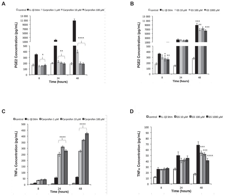
The use of dietary supplements as an alternative treatment for joint-related pathologies such as osteoarthritis (OA) is increasing. However, there is little scientific evidence to support the intended use. The aim of this study was to evaluate the anti-inflammatory effects of creatine- and amino acid-based supplements in primary cultured canine chondrocytes (CnCs) as an in-vitro model of OA and compare the effects to more commonly used agents, such as the non-steroidal anti-inflammatory drug (NSAID), carprofen, and the joint supplement, glucosamine (GS). CnCs were stimulated with interleukin-1β (IL-1β) and the subsequent release of prostaglandin E2 (PGE2) and tumor necrosis factor alpha (TNFα) was measured using an enzyme-linked immunosorbent assay (ELISA). Changes in oxylipins were also assessed using high-performance liquid chromatography/tandem mass spectrometry (HPLC/MS/MS). All compounds examined were able to significantly reduce the release of PGE2 and TNFα and were associated with reductions in cyclooxygenase-2 (COX-2) expression and nuclear factor-kappaB (NF-κB) phosphorylation. The creatine- and amino acids-based supplements also altered the profile of oxylipins produced. All compounds examined were less effective at reducing the release of PGE2 than carprofen. Carprofen significantly increased release of TNFα from CnCs, however, while the other agents reduced TNFα release. This study suggests that creatine- and amino acid-based supplements may have a beneficial role in preventing inflammation within the joint and that further studies are warranted.
L’utilisation de suppléments alimentaires à titre de traitement alternatif pour les pathologies associées aux articulations telle que l’arthrose (OA) est en augmentation. Toutefois, il y a peu d’évidences scientifiques qui supportent l’utilisation proposée. L’objectif de la présente étude était d’évaluer les effets anti-inflammatoires de suppléments à base de créatine et d’acides aminés sur des cultures primaires de chondrocytes canins (CnCs) utilisés comme modèle in vitro d’OA et de comparer les effets à des agents plus communément utilisés, tel que l’agent anti-inflammatoire non-stéroïdien (AINS) carprofen, et le supplément articulaire, glucosamine (GS). Les CnCs furent stimulés avec de l’interleukine-1β (IL-1β) et la libération subséquente de prostaglandine E2 (PGE2) et le facteur nécrosant de tumeur alpha (TNFα) fut mesurée par épreuve immuno-enzymatique (ELISA). Les changements dans les oxylipines furent également mesurés par chromatographie en phase liquide à haute performance/spectrométrie de masse tandem (HPLC/MS/MS). Tous les composés examinés étaient en mesure de réduire significativement la libération de PGE2 et de TNFα et étaient associés avec des réductions d’expression de cyclooxygénase-2 (COX-2) et de phosphorylation du facteur nucléaire kappaB (NF-κB). Les suppléments à base de créatine et d’acides aminés ont également altéré le profil des oxylipines produits. Tous les composés examinés étaient moins efficaces que le carprofen pour réduire la libération de PGE2. Le carprofen augmentait significativement la libération de TNFα par les CnCs, alors que les autres agents la réduisaient. La présente étude suggère que les suppléments à base de créatine et d’acides aminés pourraient avoir un rôle bénéfique dans la prévention de l’inflammation dans l’articulation et que des études supplémentaires sont requises.(Traduit par Docteur Serge Messier).
Figures


Similar articles
-
Pharmacological effects of a C-phycocyanin-based multicomponent nutraceutical in an in-vitro canine chondrocyte model of osteoarthritis.Can J Vet Res. 2015 Jul;79(3):241-9. Can J Vet Res. 2015. PMID: 26130858 Free PMC article.
-
Polyunsaturated fatty acids influence inflammatory markers in a cellular model for canine osteoarthritis.J Anim Physiol Anim Nutr (Berl). 2018 Apr;102(2):e623-e632. doi: 10.1111/jpn.12804. Epub 2017 Oct 13. J Anim Physiol Anim Nutr (Berl). 2018. PMID: 29030883
-
Effects of low molecular weight hyaluronan combined with carprofen on canine osteoarthritis articular chondrocytes and cartilage explants in vitro.In Vitro Cell Dev Biol Anim. 2015 Sep;51(8):857-65. doi: 10.1007/s11626-015-9908-9. Epub 2015 May 16. In Vitro Cell Dev Biol Anim. 2015. PMID: 25982358
-
Efficacy of meloxicam compared with carprofen for treating canine osteoarthritis.Vet Rec. 2020 Jan 25;186(3):94. doi: 10.1136/vr.m50. Vet Rec. 2020. PMID: 31974182 Review. No abstract available.
-
Pharmaceutical and nutraceutical management of canine osteoarthritis: present and future perspectives.Vet J. 2005 Jul;170(1):113-23. doi: 10.1016/j.tvjl.2004.08.014. Vet J. 2005. PMID: 15993795 Review.
Cited by
-
The effect of exosomes from canine bone mesenchymal stem cells on IL-1β-mediated inflammatory responses in chondrocytes.Cytotechnology. 2025 Feb;77(1):27. doi: 10.1007/s10616-024-00685-4. Epub 2024 Dec 28. Cytotechnology. 2025. PMID: 39736844
References
-
- Rychel JK. Diagnosis and treatment of osteoarthritis. Top Companion Anim Med. 2010;25:20–25. - PubMed
-
- Lane NE, Schinitzer TJ. Osteoarthritis. In: Goldman L, Schafer IA, editors. Goldman’s Cecil Medicine. 24th ed. Philadelphia, Pennsylvania: Saunders Elsevier; 2011. pp. 1672–1676.
MeSH terms
Substances
LinkOut - more resources
Full Text Sources
Medical
Research Materials