

Quick Leukocyte Nucleus Segmentation in Leukocyte Counting
- PMID: 31308855
- PMCID: PMC6594331
- DOI: 10.1155/2019/3072498
Quick Leukocyte Nucleus Segmentation in Leukocyte Counting
Abstract
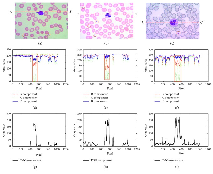
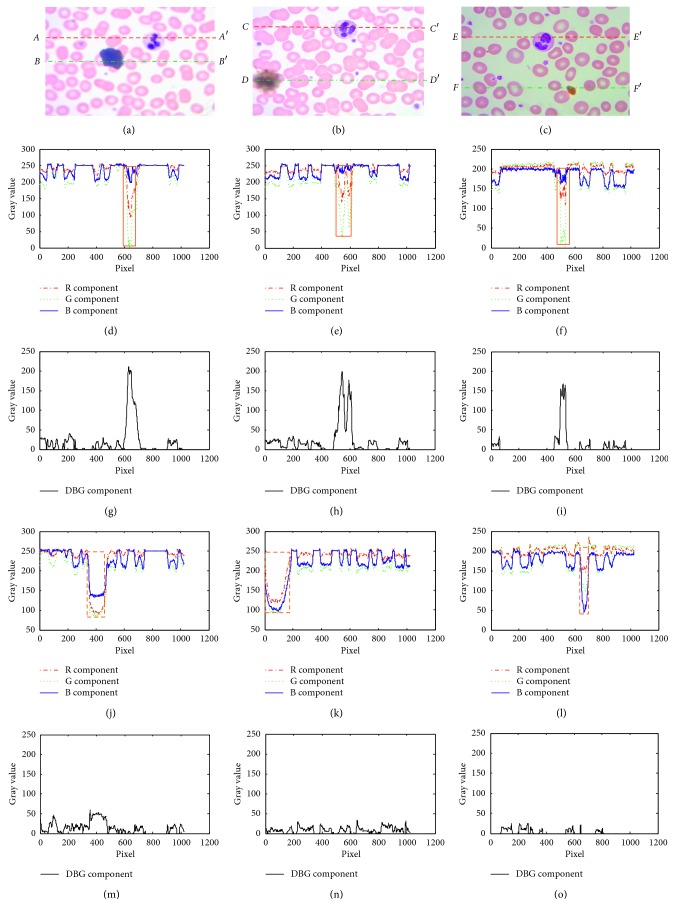
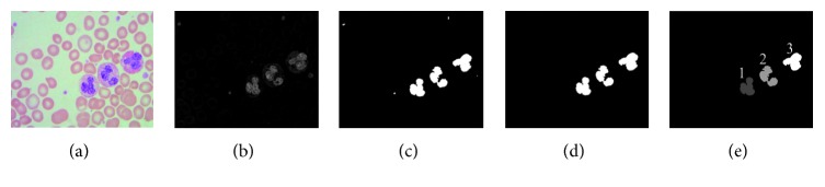
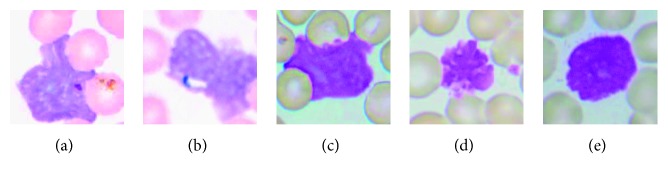
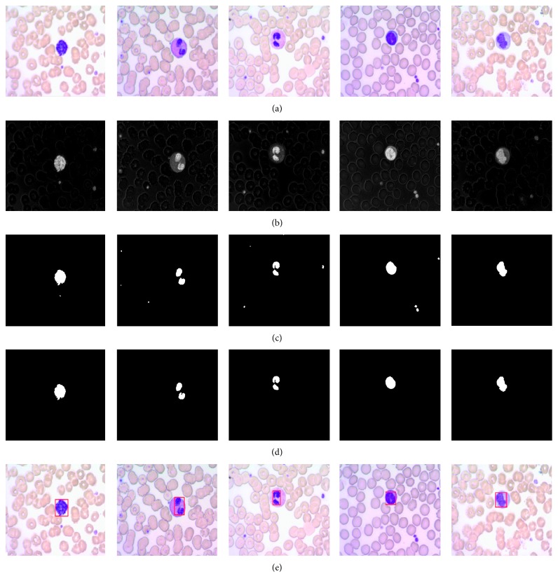
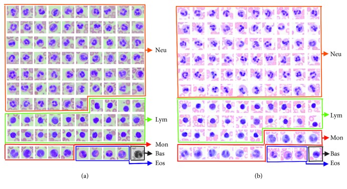
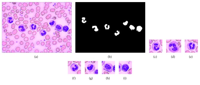
The leukocyte nucleus quick segmentation is one of the key techniques in leukocyte real-time online scanning of human blood smear. We propose a quick leukocyte nucleus segmentation method based on the component difference in RGB color space. By analyzing the captured microscopic images of the peripheral blood smears from the autoscanning microscope, it is found that the difference values between B component and G component (B - G values) in the regions of the leukocyte nuclei and the platelets are much bigger than those in the other regions, even in the regions including the stains. So, the B - G values can segment the leukocyte nuclei and the platelets with an appropriate empirical threshold because the platelets are much smaller than the leukocyte nuclei, so the leukocyte nuclei can be segmented by size filtering. Also, only an 8 bit subtraction operation is performed for the B - G values, and it can improve the leukocyte nucleus segmentation speed significantly. Experimental results show that the proposed method performs well for the five types of leukocyte segmentation with a quick speed. It is very suitable for the real-time peripheral blood smear autoscanning test application. In addition, the five types of leukocytes can be counted accurately.
Figures
Similar articles
-
Leukocyte nucleus segmentation and nucleus lobe counting.BMC Bioinformatics. 2010 Nov 12;11:558. doi: 10.1186/1471-2105-11-558. BMC Bioinformatics. 2010. PMID: 21073711 Free PMC article.
-
Image Processing Approach for Detection of Leukocytes in Peripheral Blood Smears.J Med Syst. 2019 Mar 22;43(5):114. doi: 10.1007/s10916-019-1219-3. J Med Syst. 2019. PMID: 30903283
-
Recent computational methods for white blood cell nuclei segmentation: A comparative study.Comput Methods Programs Biomed. 2019 May;173:1-14. doi: 10.1016/j.cmpb.2019.03.001. Epub 2019 Mar 6. Comput Methods Programs Biomed. 2019. PMID: 31046984 Review.
-
Segmentation of white blood cells and comparison of cell morphology by linear and naïve Bayes classifiers.Biomed Eng Online. 2015 Jun 30;14:63. doi: 10.1186/s12938-015-0037-1. Biomed Eng Online. 2015. PMID: 26123131 Free PMC article.
-
Segmentation of White Blood Cell, Nucleus and Cytoplasm in Digital Haematology Microscope Images: A Review-Challenges, Current and Future Potential Techniques.IEEE Rev Biomed Eng. 2021;14:290-306. doi: 10.1109/RBME.2020.3004639. Epub 2021 Jan 22. IEEE Rev Biomed Eng. 2021. PMID: 32746365 Review.
Cited by
-
Application of image recognition technology in pathological diagnosis of blood smears.Clin Exp Med. 2024 Aug 6;24(1):181. doi: 10.1007/s10238-024-01379-z. Clin Exp Med. 2024. PMID: 39105953 Free PMC article. Review.
-
A Polylobar Nucleus Identifying and Extracting Method for Leukocyte Counting.Comput Math Methods Med. 2021 Jul 22;2021:5565156. doi: 10.1155/2021/5565156. eCollection 2021. Comput Math Methods Med. 2021. PMID: 34335863 Free PMC article.
References
-
- Mohapatra S., Patra D., Satpathy S. An ensemble classifier system for early diagnosis of acute lymphoblastic leukemia in blood microscopic images. Neural Computing and Applications. 2014;24(7-8):1887–1904. doi: 10.1007/s00521-013-1438-3. - DOI
-
- Li X., Cao Y. A robust automatic leukocyte recognition method based on island-clustering texture. Journal of Innovative Optical Health Sciences. 2016;9(1) doi: 10.1142/s1793545816500097.1650009 - DOI
MeSH terms
LinkOut - more resources
Full Text Sources
