

Study of Surface Structure Changes for Selected Ceramics Used in the CAD/CAM System on the Degree of Microbial Colonization, In Vitro Tests
- PMID: 31309119
- PMCID: PMC6594334
- DOI: 10.1155/2019/9130806
Study of Surface Structure Changes for Selected Ceramics Used in the CAD/CAM System on the Degree of Microbial Colonization, In Vitro Tests
Abstract
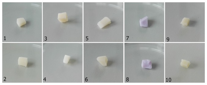
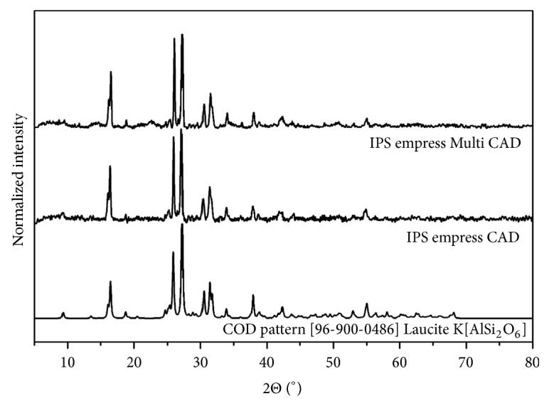
In the article has been presented an analysis of susceptibility of selected dental materials, made in the CAD/CAM technology. The morphology and structural properties of selected dental materials and their composites were determined by using XRPD (X-ray powder diffraction) techniques, as well as the IR (infrared) spectroscopy. Moreover, an adhesion as well as development of biofilm by oral microorganisms has been studied. It has been shown that a degree of the biofilm development on the tested dental materials depended on microorganism genus and species. Streptococcus mutans has demonstrated the best adhesion to the tested materials in comparison with Candida albicans and Lactobacillus rhamnosus. However, the sintered materials such as IPS e.max® and the polished IPS e.max® have showed the best "anti-adhesive properties" in relation to S. mutans and L. rhamnosus that have not formed the biofilm on the polished IPS e.max® sample. Furthermore, S. mutans have not formed the biofilm on both surfaces. On the contrary to S. mutans and L. rhamnosus, C. albicans has demonstrated the adhesive properties in relation to the above-mentioned surfaces. Moreover, in contrast to S. mutans and C. albicans, L. rhamnosus has not formed the biofilm on the polished IPS Empress material.
Figures


Similar articles
-
Interactions between Lactobacillus rhamnosus GG and oral micro-organisms in an in vitro biofilm model.BMC Microbiol. 2016 Jul 12;16(1):149. doi: 10.1186/s12866-016-0759-7. BMC Microbiol. 2016. PMID: 27405227 Free PMC article.
-
Effect of Type II Diabetes Mellitus, Candida Albicans and Streptococcus Mutans on the Biofilm Formation on Prosthetic Materials.J Contemp Dent Pract. 2018 Dec 1;19(12):1538-1545. J Contemp Dent Pract. 2018. PMID: 30713186
-
Effect of dental restorative materials surface roughness on the in vitro biofilm formation of Streptococcus mutans biofilm.Am J Dent. 2020 Apr;33(2):59-63. Am J Dent. 2020. PMID: 32259408
-
In vitro study of biofilm formation and effectiveness of antimicrobial treatment on various dental material surfaces.Mol Oral Microbiol. 2010 Dec;25(6):384-90. doi: 10.1111/j.2041-1014.2010.00586.x. Mol Oral Microbiol. 2010. PMID: 21040512
-
Effect of histatin-5 and lysozyme on the ability of Streptococcus mutans to form biofilms in in vitro conditions.Postepy Hig Med Dosw (Online). 2015 Sep 20;69:1056-66. Postepy Hig Med Dosw (Online). 2015. PMID: 26400891 Review.
Cited by
-
Key Properties of a Bioactive Ag-SiO2/TiO2 Coating on NiTi Shape Memory Alloy as Necessary at the Development of a New Class of Biomedical Materials.Int J Mol Sci. 2021 Jan 6;22(2):507. doi: 10.3390/ijms22020507. Int J Mol Sci. 2021. PMID: 33419163 Free PMC article.
-
Review on Polymer, Ceramic and Composite Materials for CAD/CAM Indirect Restorations in Dentistry-Application, Mechanical Characteristics and Comparison.Materials (Basel). 2021 Mar 24;14(7):1592. doi: 10.3390/ma14071592. Materials (Basel). 2021. PMID: 33805176 Free PMC article. Review.
-
Bacterial adhesion to composite resins produced by additive and subtractive manufacturing.Odontology. 2024 Apr;112(2):460-471. doi: 10.1007/s10266-023-00862-5. Epub 2023 Oct 11. Odontology. 2024. PMID: 37819468
-
The Influence of Ozonated Olive Oil-Loaded and Copper-Doped Nanohydroxyapatites on Planktonic Forms of Microorganisms.Nanomaterials (Basel). 2020 Oct 10;10(10):1997. doi: 10.3390/nano10101997. Nanomaterials (Basel). 2020. PMID: 33050423 Free PMC article.
-
Bacteriological Evaluation of Gingival Crevicular Fluid in Teeth Restored Using Fixed Dental Prostheses: An In Vivo Study.Int J Mol Sci. 2021 May 22;22(11):5463. doi: 10.3390/ijms22115463. Int J Mol Sci. 2021. PMID: 34067261 Free PMC article.
References
-
- Lebon N., Tapie L., Duret F., Attal J.-P. Understanding Dental CAD/CAM for Restorations-Dental Milling Machines from a Mechanical Engineering Viewpoint. Part B: Labside Milling Machines. International Journal of Computerized Dentistry. 2016;19(2):115–134. - PubMed
-
- Teughels W., Van Assche N., Sliepen I., Quirynen M. Effect of material characteristics and/or surface topography on biofilm development. Clinical Oral Implants Research. 2006;17, Supplement 2:68–81. - PubMed
-
- Gopikrishna V. Preclinical Manual of Conservative Dentistry and Endodontics. 2nd. India: Elsevier; 2015.
MeSH terms
Substances
LinkOut - more resources
Full Text Sources
Molecular Biology Databases
Miscellaneous
