Review
. 2020 Jun;41(Suppl 1):9-19.
doi: 10.1007/s00292-019-0612-5.

Complexity of PEComas : Diagnostic approach, molecular background, clinical management

Affiliations
Review

Complexity of PEComas : Diagnostic approach, molecular background, clinical management

K Utpatel et al. Pathologe. 2020 Jun.

Abstract

Perivascular epithelioid cell neoplasms (PEComas) are a family of mesenchymal neoplasms with features of both melanotic and smooth muscle differentiation. PEComa morphology is highly variable and encompasses epithelioid to spindle cells often with clear cytoplasm and prominent nucleoli. Molecularly, most PEComas are defined by a loss of function of the TSC1/TSC2 complex. Additionally, a distinct small subset of PEComas harboring rearrangements of the TFE3 (Xp11) gene locus has been identified. By presenting a series of three case reports with distinct features, we demonstrate diagnostic pitfalls as well as the importance of molecular work-up of PEComas because of important therapeutic consequences.

Perivaskuläre epitheloidzellige Tumoren (PECome) gehören zu einer Familie von mesenchymalen Neoplasien mit Merkmalen der melanotischen und glattmuskulären Differenzierung. Die Morphologie der PECome ist sehr variabel und umfasst epitheloide und spindelige Zellen, oft mit klarem Zytoplasma und prominenten Nukleoli. Molekular sind die meisten PECome durch einen Funktionsverlust des TSC1-TSC2-Komplexes definiert. Zusätzlich wurde eine kleine Untergruppe von PEComen identifiziert, die Rearrangements des TFE3(Xp11)-Genlocus aufzeigen. Anhand von 3 Fallberichten sollen die diagnostischen Fallstricke und die Bedeutung der molekularen Charakterisierung von PEComen auch wegen der therapeutischen Konsequenzen näher dargestellt werden.

Keywords: Genetic translocation; Immunohistochemistry; Lymphangioleiomyomatosis; Perivascular epithelioid cell neoplasms; TOR serine-threonine kinases.


Conflict of interest statement

K. Utpatel, D.F. Calvisi, G. Köhler, T. Kühnel, A. Niesel, N. Verloh, M. Vogelhuber, R. Neu, N. Hosten, H.-U. Schildhaus, W. Dietmaier, and M. Evert declare that they have no competing interests.

Figures

Fig. 1
Fig. 1
Case 1: Histological and immunohistochemical aspects. a H&E, ×200; b HMB-45, ×100; c caldesmon, ×100; d p-mTOR, ×100; e p-4EBP1, ×100; f p-RPS6, ×100
Fig. 2
Fig. 2
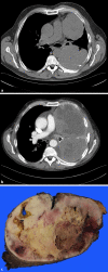
Case 1: Computed tomography scan of the thoracic relapsed tumor and macroscopy of the resected tumor. a Two new tumor nodules in the left thorax in July 2018. b Tumor progression in November 2018. c Resected tumor after 5‑month mTOR inhibitor treatment
Fig. 3
Fig. 3
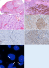
Case 2: Melanotic perivascular epithelioid cell neoplasm of the nasal cavity. a Overview, H&E, ×16; b detection of melanin, ×200; c Atypical tumor cells, ×100; d HMB-45, ×100×; e S-100, ×100; f TFE3, ×100; g fluorescence in situ hybridization: TFE3 break-apart probe reveals small gaps between 3′TFE3 and 5′ TFE3 in 3 interphase cells (arrows) in a male patient, indicating the translocation of the only X chromosome
Fig. 4
Fig. 4
Case 3: Contrast-enhanced computer tomography before everolimus treatment and a follow-up scan 4 months after initial treatment. a Large tumor detected on the right side in the pelvis (black arrows); b lung metastasis (white arrow); c detection of a small scarred residual tumor 4 months after treatment (black arrow); d no lung metastasis can be observed
Fig. 5
Fig. 5
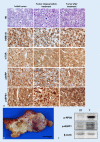
Case 3: Malignant perivascular epithelioid cell neoplasm of the uterus. a H&E, HMB-45, p‑mTOR, p‑4EBP1, and p‑RPS6, ×400. b Macroscopy of the residual pelvic tumor after 4 months of mTOR inhibitor treatment. c Western blot analysis of mTOR downstream effectors p‑RPS6 and p‑4EBP1 in the tumor (T) and non-tumorous surrounding tissue (ST). β‑Actin was used as a loading control. Strong induction of the two mTOR effector proteins in the tumor part
Fig. 6
Fig. 6
Scheme depicting the two main molecular subgroups of perivascular epithelioid cell neoplasms (PEComa) identified to date. In the first subtype, the most frequent type, loss of function of the complex consisting of TSC Complex Subunit 1 and 2 (TSC1 and TSC2) proteins leads to activation of the Ras homolog, mTORC 1 binding protein (RHEB), with consequent induction of mTORC1. Once activated, mTORC1 triggers the activation of its downstream targets, S6 ribosomal protein (RPS6) as well as the inhibition of the tumor suppressor 4EBP1, thus leading to unrestrained cell growth. mTORC1-driven events are mediated by phosphorylation (p) mechanisms. In the second PEComa molecular subtype, various molecular mechanisms (amplifications, gene fusion, etc.) induce elevated transcriptional activity of TFE3, resulting in activation of the c‑Met proto-oncogene and induction of the downstream effector proteins, which are partly known (AKT, mTOR) and partly unknown (???), harboring important pro-oncogenic and growth properties. Of note, many of the inducers and effectors described can be specifically inhibited (as indicated by red, blunted arrows) by available drugs

References

    1. Albores-Saavedra J, Dorantes-Heredia R, Chable-Montero F, et al. Endometrial stromal sarcomas: immunoprofile with emphasis on HMB45 reactivity. Am J Clin Pathol. 2014;141:850–855. doi: 10.1309/AJCPS88CMJRXZBWA. - DOI - PubMed
    1. Argani P, Aulmann S, Illei PB, et al. A distinctive subset of PEComas harbors TFE3 gene fusions. Am J Surg Pathol. 2010;34:1395–1406. doi: 10.1097/PAS.0b013e3181f17ac0. - DOI - PubMed
    1. Argani P, Lal P, Hutchinson B, et al. Aberrant nuclear immunoreactivity for TFE3 in neoplasms with TFE3 gene fusions: a sensitive and specific immunohistochemical assay. Am J Surg Pathol. 2003;27:750–761. doi: 10.1097/00000478-200306000-00005. - DOI - PubMed
    1. Argani P, Zhong M, Reuter VE, et al. TFE3-fusion variant analysis defines specific clinicopathologic associations among Xp11 translocation cancers. Am J Surg Pathol. 2016;40:723–737. doi: 10.1097/PAS.0000000000000631. - DOI - PMC - PubMed
    1. Au KS, Williams AT, Roach ES, et al. Genotype/phenotype correlation in 325 individuals referred for a diagnosis of tuberous sclerosis complex in the United States. Genet Med Off J Am Coll Med Genet. 2007;9:88–100. - PubMed

MeSH terms

LinkOut - more resources