Review
. 2019 Jul;25(7):e3198.
doi: 10.1002/psc.3198.

Templated chemistry for bioorganic synthesis and chemical biology

Affiliations
Review

Templated chemistry for bioorganic synthesis and chemical biology

Oliver Seitz. J Pept Sci. 2019 Jul.

Abstract

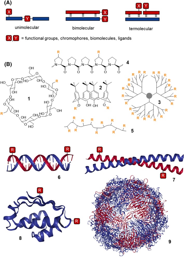
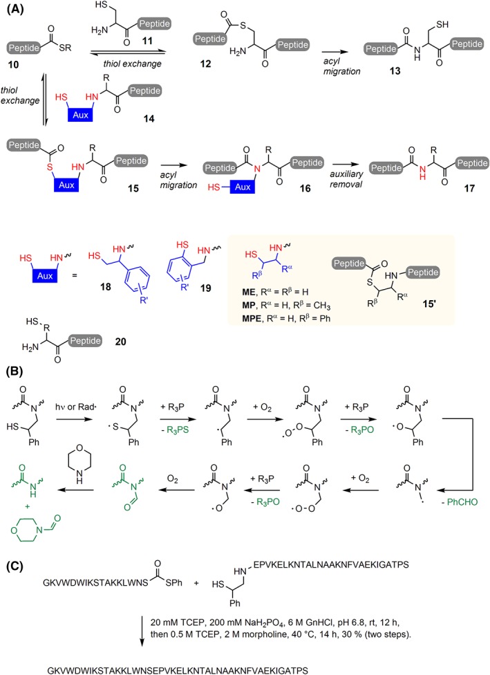
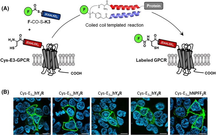
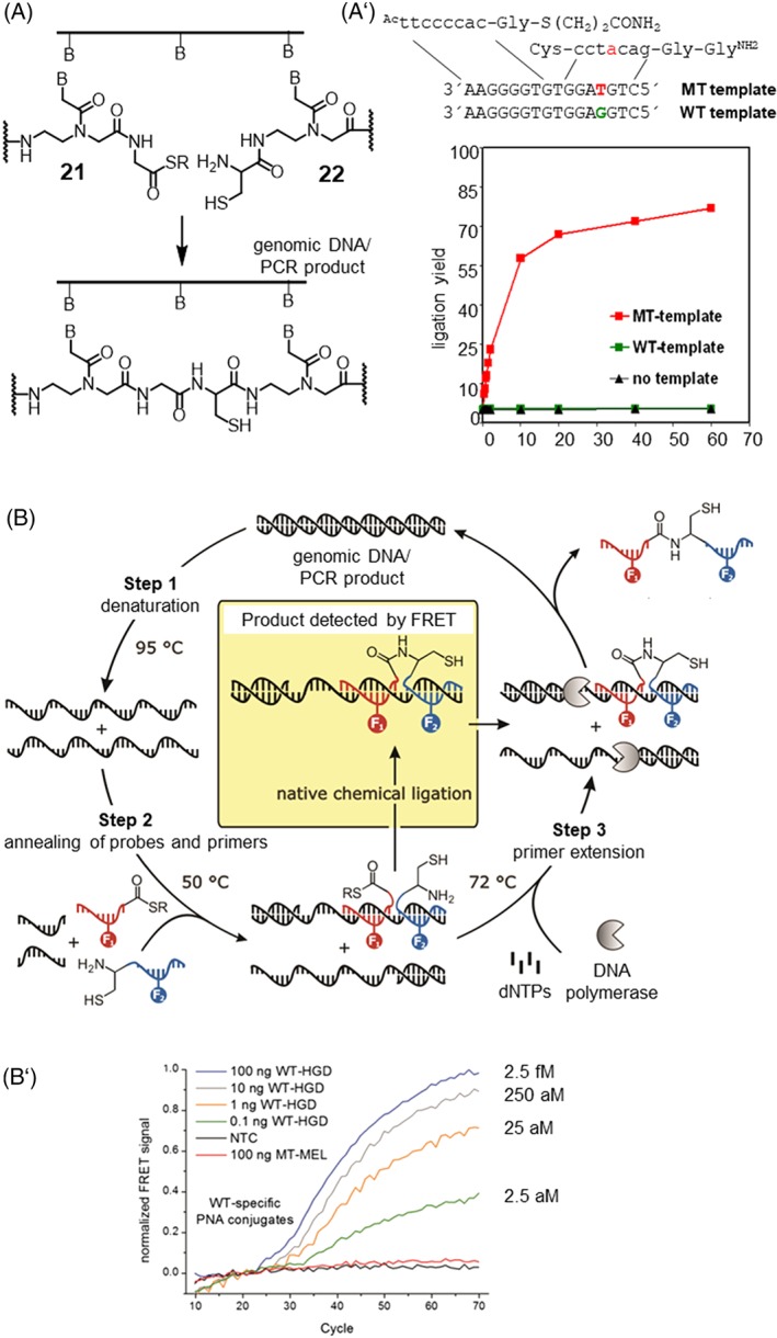
In light of the 2018 Max Bergmann Medal, this review discusses advancements on chemical biology-driven templated chemistry developed in the author's laboratories. The focused review introduces the template categories applied to orient functional units such as functional groups, chromophores, biomolecules, or ligands in space. Unimolecular templates applied in protein synthesis facilitate fragment coupling of unprotected peptides. Templating via bimolecular assemblies provides control over proximity relationships between functional units of two molecules. As an instructive example, the coiled coil peptide-templated labelling of receptor proteins on live cells will be shown. Termolecular assemblies provide the opportunity to put the proximity of functional units on two (bio)molecules under the control of a third party molecule. This allows the design of conditional bimolecular reactions. A notable example is DNA/RNA-triggered peptide synthesis. The last section shows how termolecular and multimolecular assemblies can be used to better characterize and understand multivalent protein-ligand interactions.

Keywords: Oligonucleotides; bioconjugation; carbohydrates; lectin; multivalency; peptide ligation; protein labeling; templated synthesis.


Figures

Figure 1
Figure 1
A, Templating strategies to arrange the functional units X and Y in defined orientation and distance. B, Representative examples of template molecules used for presentation of groups R (PDB IDs: 6, 1LAI; 7, 2XU6; 8, 1BTA; 9, 2VTU)
Figure 2
Figure 2
A, Native chemical ligation (10 + 11) and auxiliary mediated native chemical ligation (10 + 14). B, Proposed radical‐induced oxidative fragmentation of the MPE auxiliary. Products highlighted in green were detected by high‐performance liquid chromatography–mass spectrometry (HPLC‐MS) and nuclear magnetic resonance (NMR). C, One‐pot ligation auxiliary removal in the synthesis of opistoporin‐2 (GnHCl, guanidinium hydrochloride; TCEP, triscarboxyethylphosphine)
Figure 3
Figure 3
A, Formation of the E3‐K3 coiled coil allows the rapid and selective labelling of a Cys‐E3 tagged GPCR by thioester‐linked fluorophore‐K3 peptide conjugates. B, Live cell fluorescence microscopy images after 5‐min incubation of transiently transfected HEK293 cells (Cys‐E3_hY1R, Cys‐E3_hY2R, Cys‐E3_hY4R, Cys‐E3_hY5R, and Cys‐E3_hNPFF2R) with 100 nM ATTO488‐K3 conjugate (reproduced from Reinhardt et al68 copyright 2015 American Chemical Society)
Figure 4
Figure 4
A, Principle of nucleic acid–templated native chemical ligation (NCL) of peptide nucleic acid (PNA). A′, Reaction time course of templated PNA NCL on matched and mismatched template (conditions: 1 μM PNA probes and templates, 100 mM Na2HPO4, sat BnSH, pH 7.4, 25°C). B, DNA template NCL during PCR (reproduced from Roloff and Seitz90 with permission from The Royal Society of Chemistry). B′, Amplification plots obtained by determining the normalized fluorescence resonance energy transfer Föster resonance energy transfer (FRET) signal upon ligation during polymerase chain reaction (PCR) in the presence of varying amounts of matched (Raf‐wt human genomic DNA, WT‐HGD) or mismatched (Raf‐T1799A‐mt human genomic DNA, MT‐HGD) DNA target (PCR conditions: 400 nM forward primer, 50 nM reverse primer, 200 mM dNTPs, 2.5 mM MgCl2, 300 nM PNA conjugates, 1 mM MESNA, 10 mM TRIS, pH 8.5 (at 25°C), 1 u Taq‐Pol; PCR protocol: 10 s at 95°C (step 1), 30 s at 50°C (step 2, detection), 20 s at 72°C (step 3))
Figure 5
Figure 5
A, Nucleic acid–templated acyl transfer reaction. B, Reporter groups used in templated acyl transfer reactions. C, Yield of transfer reaction between FAM‐AEEA‐tcttccccac‐Cys (Gly‐Dabcyl) and iCys‐cctacag‐Lys (AEEA‐TAMRA) at substoichiometric amounts of template 5′‐GCCGCTGTAGGTGTGGGGAAGAGT‐3′ (conditions: 100 nM probes, 10 mM NaH2PO4, 200 mM NaCl, 1 mM triscarboxyethylphosphine (TCEP), 0.1 mg/mL Roche blocking agent, pH 7.0, 32°C)
Figure 6
Figure 6
A, Templated alanyl transfer induces the formation of peptide 29, which displaces caspase‐9 from the XIAP‐BIR3 domain. B, Relative activity of caspase‐9 and caspase‐3 in HEK293 cell lysate (L) and after addition of the XIAP‐BIR3 domain (L + BIR3). Addition of probes 27 and 28 in the presence of matched template (Ma) led to caspase reactivation. No reactivation was observed in the absence of template (BG), with mismatched template (Mi) or in template only experiments (only Ma) (reproduced from Erben et al107; copyright 2011 Wiley)
Figure 7
Figure 7
A, RNA‐templated peptidyl transfer reaction between 30 and 31 leads to conjugate 32, which is cyctoxic when produced in excess to template. B, Inhibition of HeLa cell proliferation (MTS assay) after treatment with reactive probes in the presence of matched (XIAP‐RNA) and mismatched (GAPDH‐RNA) template (reproduced from Vazquez and Seitz108 with permission from The Royal Society of Chemistry)
Figure 8
Figure 8
A, DNA‐templated self‐assembly with nucleic acid–ligand conjugates provides control over the valency and spatial arrangement of ligand displays. B, DNA‐programmed spatial screening of interactions between bivalent displays of LacNAc and Erythrina cristagalli lectin (ECL, top) or Ricinus communis agglutinin (RCA120, bottom) reveals distinct distance‐affinity relationships
Figure 9
Figure 9
A, Crystal structures of Syk and Zap‐70 tSH2 domains in complex with double phosphorylated immunoreceptor tyrosine–based interaction motifs (ITAMs) from the CD3ε chain of the B‐cell receptor complex or the ζ‐chain of the T‐cell receptor, respectively (from PDB ID 1A81 and 2OQ1, reproduced from Marczynke et al121; copyright 2017 American Chemical Society). B, DNA‐peptide complexes used for DNA‐programmed spatial screening. Distance‐affinity relationships for interactions with Syk tSH2 (gray) or Zap‐70 tSH2 (black) obtained by spatial screening with complexes displaying peptides in C, same strand orientation or D, opposing strand orientation
Figure 10
Figure 10
A, Relative binding affinity (RBA, relative to estradiol) of bivalent raloxifene displays for the estrogen receptor α. Nonconjugated raloxifene has RBA = 30%. B, Docking (reproduced with permission from Abendroth et al124; copyright 2011 Wiley) of Ral 2 ‐DNA_3 (raloxifene shown in green) to the ERα ligand binding domain with the depiction of unconjugated raloxifene (magenta) in a cocrystal structure (PDB ID: 2R6W)
Figure 11
Figure 11
A, Cryo‐electron micrograph of human influenza virus (X31) and depiction of trimeric hemagglutinin (HA) with position of canonical sugar binding sites marked in yellow. The distance between two binding sites within a single HA trimer was extracted from crystal structure analysis (PDB ID: 1HGG, reproduced from Bandlow et al129; copyright 2017 American Chemical Society). The distances between two adjacent HA trimers were determined by cryo–transmission electron microscopy (cryo‐TEM) analysis. Given free rotation of HA trimers, two canonical glyco binding sites on adjacent HA trimers could be arranged in 49 to 154 Å distance. B, Bivalent displays for probing of intertrimeric and intratrimeric HA interactions. C, Distance‐dependent inhibition of influenza X31 hemagglutination by bivalent Sialyl‐LacNAc conjugates. Open squares show data for the highest concentration of conjugate applied at which hemagglutination was still not inhibited (reproduced from Bandlow et al129; copyright 2017 American Chemical Society). D, Concatenation of optimized bivalent Sialyl‐LacNAc displays by linear (top) or branched (bottom) hybridization with long DNA templates produced by rolling circle amplification. The values KiHAI are based on the concentration of sugar (SLN) or template required to fully inhibit hemagglutination. E, Cryo‐electron micrograph of DNA‐concatenated SLN incubated with IAV X31 for 30 min and embedded in vitreous ice. Linear spaghetti‐type structures are highlighted in red and orange arrows. F, Slice of the reconstructed 3D volume of tilt series showing a virus‐bound wool‐type SLN concatemer. Scale bars: 50 nm. E and F reproduced from Bandlow et al130; copyright 2019 Wiley
Figure 12
Figure 12
A, Graphical presentation of the heterotetrameric adaptor protein AP‐2 in complex with peptides that bind to the top and side sites of the ear domains (model based on PDB IDs 2VGL, 2VJO, 2G30, and 3HS9, modified from Dubel et al137; copyright 2019 Wiley). B, Representative examples of cucurbituril CB7– and adamantane‐DNA conjugates used to probe the distance‐stability relationship of complexes formed upon bivalent interaction. C, Distance dependency of bivalency‐enhanced interactions between distance‐matched CB7 and adamantane displays (bivalency enhancement = K d (mono)/2·K d (biv))

Similar articles

Cited by

References

    1. Prescher JA, Bertozzi CR. Chemistry in living systems. Nat Chem Biol. 2005;1(1):13‐21. - PubMed
    1. Johnsson N, Johnsson K. Chemical tools for biomolecular imaging. ACS Chem Biol. 2007;2(1):31‐38. - PubMed
    1. Salic A, Mitchison TJ. A chemical method for fast and sensitive detection of DNA synthesis in vivo. Proc Natl Acad Sci U S a. 2008;105(7):2415‐2420. - PMC - PubMed
    1. Jao CY, Salic A. Exploring RNA transcription and turnover in vivo by using click chemistry. Proc Natl Acad Sci U S a. 2008;105(41):15779‐15784. - PMC - PubMed
    1. Holstein JM, Rentmeister A. Current covalent modification methods for detecting RNA in fixed and living cells. Methods. 2016;98:18‐25. - PubMed

LinkOut - more resources