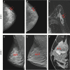
[Digital breast tomosynthesis in diagnosis of dense breast lesions]
- PMID: 31309757
- PMCID: PMC8800639
- DOI: 10.3785/j.issn.1008-9292.2019.04.10
[Digital breast tomosynthesis in diagnosis of dense breast lesions]
Abstract
Objective: To evaluate the value of digital breast tomosynthesis (DBT) in diagnosis of dense breast lesions.
Methods: Clinical and pathological data of 163 patients (58 benign lesions, 122 malignant lesions, and 180 lesions in total) with breast lesions undergoing surgical treatment in Shaoxing Central Hospital from January 2017 to December 2018 were retrospectively analyzed. The lesions were classified into non-homogeneous dense gland type and extremely dense gland type according to BI-RADS creterion. Breast MRI and DBT examinations were performed before the surgery. ROC curve was generated and the diagnostic efficacy of two examination methods for dense breast lesions was evaluated with pathological results as the gold standard. The detection rate, diagnostic accuracy of benign and malignant breast lesions were compared between two methods using chi-square test. The accuracy of lesion size preoperatively evaluated by MRI and DBT was analyzed by Pearson correlation.
Results: The detection rate and diagnostic accuracy for benign breast lesions by MRI were higher than those by DBT (91.4% vs. 75.9%, χ2=5.098, P<0.05 and 89.7% vs. 67.2%, χ2=8.617, P<0.01). But there were no significant differences in detection rate and accuracy for malignant lesions by MRI and DBT (98.4% vs. 95.1%, χ2=2.068, P>0.05 and 94.3% vs. 91.8%, χ2=0.569, P>0.05). The areas under the ROC curves of MRI, DBT based on BI-RADS classification were 0.910 and 0.832, respectively (Z=1.860, P>0.05). The sensitivities of MRI, DBT to breast lesions were 93.3% and 86.7%, and the specificities were 68.3% and 79.1%. DBT and MRI measurements were positively correlated with pathological measurements (r=0.887 and 0.949, all P<0.01).
Conclusions: DBT can effectively diagnose benign and malignant breast lesions under dense gland background, and it has similar diagnostic efficacy with MRI for breast malignant lesions.
目的: 探讨数字化乳腺断层融合摄影(DBT)在致密型乳腺患者中诊断乳腺良恶性病变的效能。
方法: 收集2017年1月至2018年12月绍兴市中心医院经病理组织学检查证实的163例乳腺良恶性病变患者的资料(良性病灶58个,恶性病灶122个,共计180个)。根据BI-RADS标准患者乳腺腺体类型均归类于不均质致密腺体型和极度致密型。所有患者术前均行乳腺MRI、DBT检查。以病理结果为金标准绘制ROC曲线,评价两种影像学检查
方法: 对乳腺良恶性病变的诊断效能,用 Z检验对ROC曲线下面积进行比较;采用 χ 2检验比较乳腺MRI和DBT检查对乳腺良恶性病变检出率、诊断准确率的差异;采用Pearson相关性分析MRI、DBT术前评估乳腺病灶大小的准确度。
结果: MRI、DBT对乳腺良性病变的检出率和准确率分别为91.4%、75.9%和89.7%、67.2%,差异有统计学意义( χ 2=5.098、8.617, P < 0.05或 P < 0.01);MRI、DBT对乳腺恶性病变的检出率和准确率分别为98.4%、95.1%和94.3%、91.8%,差异无统计学意义( χ 2=2.068、0.569,均 P>0.05)。MRI、DBT诊断致密型乳腺病变的ROC曲线下面积分别为0.910、0.832,差异无统计学意义( Z=1.860, P>0.05)。MRI、DBT诊断致密型乳腺良恶性病变的敏感度分别为93.3%、86.7%,特异度分别为68.3%、79.1%。DBT、MRI测量值与病理测量结果呈正相关( r=0.887、0.949,均 P < 0.01)。
结论: DBT简单易行,能较好地诊断致密腺体背景下乳腺的良恶性病变,尤其对乳腺恶性病变的诊断效能与乳腺MRI相近。
Figures
Similar articles
-
[Comparison of the Diagnostic Values of Dynamic Enhanced Magnetic Resonance Imaging,Digital Breast Tomosynthesis,and Digital Mammography for Early Breast Cancer].Zhongguo Yi Xue Ke Xue Yuan Xue Bao. 2019 Oct 30;41(5):667-672. doi: 10.3881/j.issn.1000-503X.10938. Zhongguo Yi Xue Ke Xue Yuan Xue Bao. 2019. PMID: 31699198 Chinese.
-
Non-calcified ductal carcinoma in situ of the breast: comparison of diagnostic accuracy of digital breast tomosynthesis, digital mammography, and ultrasonography.Breast Cancer. 2017 Jul;24(4):562-570. doi: 10.1007/s12282-016-0739-7. Epub 2016 Nov 11. Breast Cancer. 2017. PMID: 27837442
-
The role of breast tomosynthesis in a predominantly dense breast population at a tertiary breast centre: breast density assessment and diagnostic performance in comparison with MRI.Eur Radiol. 2018 Aug;28(8):3194-3203. doi: 10.1007/s00330-017-5297-7. Epub 2018 Feb 19. Eur Radiol. 2018. PMID: 29460074 Free PMC article.
-
Digital breast tomosynthesis and contrast-enhanced dual-energy digital mammography alone and in combination compared to 2D digital synthetized mammography and MR imaging in breast cancer detection and classification.Breast J. 2020 May;26(5):860-872. doi: 10.1111/tbj.13739. Epub 2019 Dec 30. Breast J. 2020. PMID: 31886607
-
Diagnostic Performance of Adjunctive Imaging Modalities Compared to Mammography Alone in Women with Non-Dense and Dense Breasts: A Systematic Review and Meta-Analysis.Clin Breast Cancer. 2021 Aug;21(4):278-291. doi: 10.1016/j.clbc.2021.03.006. Epub 2021 Mar 16. Clin Breast Cancer. 2021. PMID: 33846098
References
-
- 边 甜甜, 林 青, 李 丽丽, et al. 对比数字乳腺断层合成与乳腺X线摄影对致密型乳腺内肿块的诊断价值. 中华放射学杂志. 2015;49(7):483–487. doi: 10.3760/cma.j.issn.1005-1201.2015.07.002. [边甜甜, 林青, 李丽丽, 等.对比数字乳腺断层合成与乳腺X线摄影对致密型乳腺内肿块的诊断价值[J].中华放射学杂志, 2015, 49(7):483-487.] - DOI
-
- DESTOUNIS S. Role of digital breast tomosynthesis in screening and diagnostic breast imaging. Semin Ultrasound CT MR. 2018;39(1):35–44. doi: 10.1053/j.sult.2017.08.002. [DESTOUNIS S. Role of digital breast tomosynthesis in screening and diagnostic breast imaging[J]. Semin Ultrasound CT MR, 2018, 39(1):35-44.] - DOI - PubMed
MeSH terms
LinkOut - more resources
Full Text Sources
Medical


