Review
. 2019 Sep 20;431(20):4007-4029.
doi: 10.1016/j.jmb.2019.07.017. Epub 2019 Jul 13.

Mechanisms of Transcriptional Pausing in Bacteria

Affiliations
Review

Mechanisms of Transcriptional Pausing in Bacteria

Jin Young Kang et al. J Mol Biol. .

Abstract

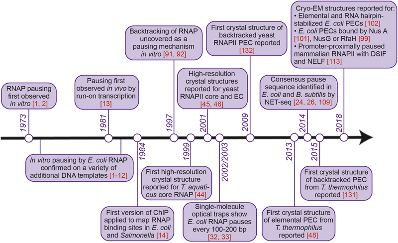
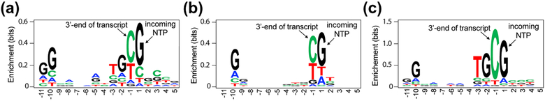
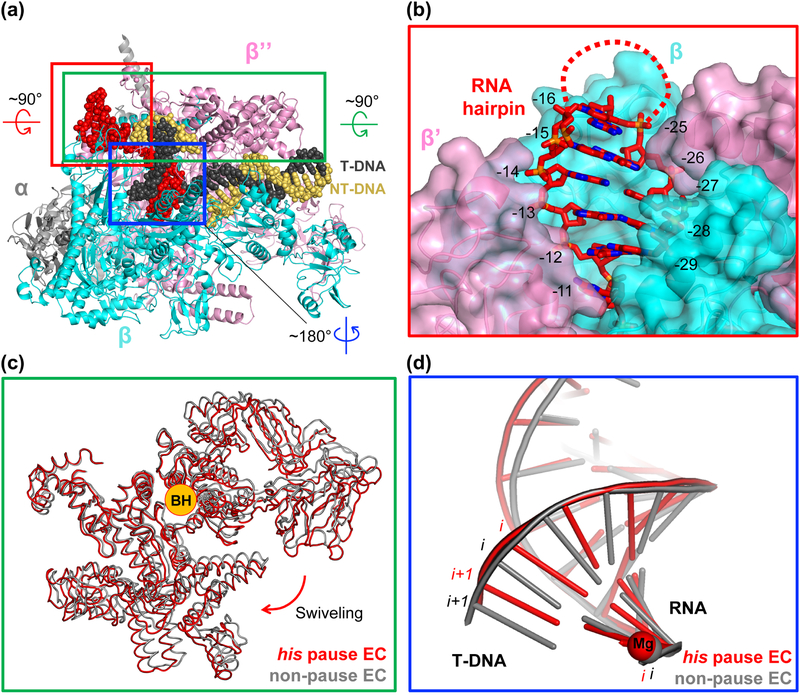
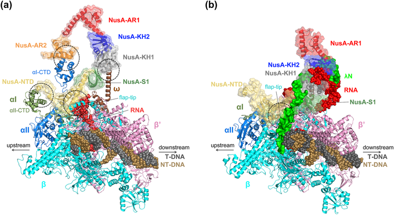
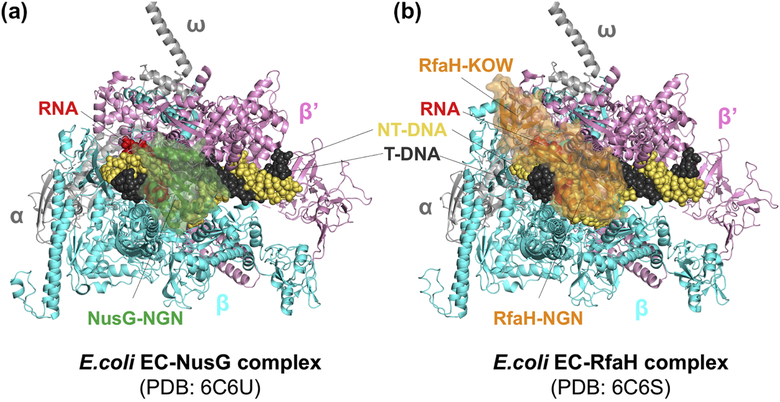
Pausing by RNA polymerase (RNAP) during transcription regulates gene expression in all domains of life. In this review, we recap the history of transcriptional pausing discovery, summarize advances in our understanding of the underlying causes of pausing since then, and describe new insights into the pausing mechanisms and pause modulation by transcription factors gained from structural and biochemical experiments. The accumulated evidence to date suggests that upon encountering a pause signal in the nucleic-acid sequence being transcribed, RNAP rearranges into an elemental, catalytically inactive conformer unable to load NTP substrate. The conformation, and as a consequence lifetime, of an elemental paused RNAP is modulated by backtracking, nascent RNA structure, binding of transcription regulators, or a combination of these mechanisms. We conclude the review by outlining open questions and directions for future research in the field of transcriptional pausing.

Keywords: Backtrack pause; Elemental pause; NusA; NusG; RNA hairpin pause.


Figures

Figure 1.
Figure 1.
Milestones in the field of transcriptional pausing research, with emphasis on pausing by bacterial RNA polymerase.
Figure 2.
Figure 2.. First high-resolution structures of RNAP.
A. First high resolution structure of a bacterial RNAP, T. aquaticus RNAP core at 3.3 Å resolution (Zhang et al., 1999). B. First high resolution structure of a eukaryotic RNAP, S. cerevisiae RNAP II core at 2.8 Å resolution (Cramer et al., 2001). All subunits are labeled on the figure and catalytic magnesium is drawn as an orange sphere in the center of the structures.
Figure 3.
Figure 3.. Schematic diagram of nucleotide addition cycle.
An active-site view of the nucleotide addition cycle by RNAP. A part of RNA-DNA hybrid in the proximity of the catalytic magnesium is drawn with the neighboring protein components, BH, TL and SI3 connected to the TL. Clamp domain of RNAP forms the main channel that accomodates the RNA-DNA hybrid, and the flap domain is located at the upstream end of RNA-DNA hybrid. The locations of clamp and flap domains are marked by colored ovals and their sizes are not to scale. Each round of nucletide addition occurs in the following steps: (i) nucleic-acid translocation from pre- to post-translocated register, which vacates the binding site for the incoming nucleoside triphosphate (NTP) and aligns the next template DNA base for base-pairing with NTP; (ii) NTP binding; (iii) TL folding into the trigger helices (TH); and (iv) formation of the phosphodiester bond. Unfolding of the TH and release of pyrophosphate accompany the translocation step (i).
Figure 4.
Figure 4.. Schematic diagram of transcriptional pauses.
In the on-pathway process of nucleotide addition, RNAP translocates from pre-translocated to post-translocated register to vacate the i+1 site for the next nucleotide to bind as shown in the top part of Figure 3. During this translocation, RNAP can isomerize into an off-pathway conformation that blocks catalysis for a few seconds in response to the nucleic acid sequence interacting with the enzyme. This isomerized state of an elongation complex (EC) is termed the ‘elemental pause’. Elemental paused EC samples multiple RNA-DNA hybrid registers, including the half-translocated register (see the main text). Elemental paused EC can further isomerize into longer-lived paused states, such as backtrack and RNA hairpin-stabilized pauses, or precede transcription termination. In an RNA hairpin-stabilized paused EC, RNA hairpin formation in the RNA exit channel induces conformational change of RNAP (swiveling), while RNA-DNA hybrid remains half-translocated. To escape a hairpin-stabilized pause, the minor population of swiveled RNAP with post-translocated RNA-DNA hybrid may be unswiveled, returning to the on-pathway. In a backtrack paused EC, RNAP moves backward on the nucleic acids, extruding the 3′-end of RNA into the secondary channel. Hybrid conformation here is drawn with dotted lines because both canonical pre-translocated and the half-translocated hybrid conformations have been observed in backtracked ECs. During intrinsic termination, RNA hairpin in a paused EC invades RNA-DNA hybrid, while Rho-dependent termination is likely preceded by an elemental pause. The conformation of RNA-DNA hybrid in the termination process is unknown.
Figure 5.
Figure 5.. Consensus elemental pause sequence.
A. Consensus sequence logo obtained by using NET-seq from B. subtilis and E. coli. Cells were harvested by filtration and flash-frozen in liquid nitrogen before RNA isolation. Native elongating transcripts were harvested by immunoprecipitating Flag-tagged RNAP and the transcripts were convered to DNA for sequencing (Larson, 2014). B. Consensus sequence obtained by NET-seq in E. coli under conditions similar to those in panel A, except the cells were not flash-frozen in liquid nitrogen but instead cooled on dry ice. (Vvedenskaya, 2014) C. Consensus sequence logo obtained by RNET-seq (read-length-specific NET-seq) (Imashimizu, 2015). Contrast to the experiments performed in above panel B and C, only the RNA region buried in RNAP was isolated and analyzed.
Figure 6.
Figure 6.. Structural features of an RNA hairpin-stabilized pause elongation complex (hisPEC).
A. The overall structure of E. coli hisPEC. Specific areas are boxed, rotated, and zoomed for detailed description. B. Red boxed region from A rotated by 90° and magnified to show RNA exit channel with his pause RNA hairpin stem duplex in it. RNA hairpin stem duplex formed fully and resided in the RNA exit channel while the loop region of the hairpin was disordered (marked by a dotted line). The opening of the channel widened compared to that of non-paused EC. The residues in RNA are numbered by their positions relative to the active site, where the RNA 3’-end nucleotide is at −1 position. The −11 RNA base connects RNA-DNA hybrid and RNA hairpin. C. Green boxed region from panel A rotated by 90° and zoomed in. Comparison of Cα traces of swivel modules from hisPEC and non-pause EC (PDB code: 6ALF). Compared to the swivel module of a non-pause EC, swivel module in hisPEC rotated about 3° around the axis overlapping BH. D. Blue boxed region from panel A rotated by 180° and zoomed in. Only nucleic acid structures were drawn for clarity. Non-pause EC exhibits a post-translocated hybrid while hisPEC has a half-translocated hybrid, harboring DNA base in i+1 site base-paired with RNA base in i site.
Figure 7.
Figure 7.. Comparison of NusA-bound hisPEC and λN anti-termination complex.
Protein region is drawn in cartoon format and nucleic acids are drawn as spheres. NusA is marked by a transparent surface and each domain of NusA is colored differently. A. hisPEC-NusA complex (PDB code: 6FLQ). The main contact points between RNAP and NusA are marked with dotted circles. B. λN anti-termination complex (PDB code: 6GOV) drawn from the same vantage point as in panel A. λN is drawn as a non-transparent green surface. The main contact point between RNAP and NusA is marked with a dotted circle. NusA-AR2 was disordered. NusB and NusE are not shown for clarity.
Figure 8.
Figure 8.. NusG- and RfaH-bound EC structures.
RNAP subunits and nucleic acids are labelled. A. E. coli NusG (colored in green) binds between β and β’ subunits, contacting β protrusion, β lobe, and β’ clamp helices. NusG KOW domain was disordered in the cryo-EM structure. B. E. coli RfaH (colored in orange) binds to the same site as NusG in panel A. In addition, RfaH recognizes a short-hairpin formed by ops sequence in the non-template DNA (not visible in the figure) and RfaH KOW domain binds flap tip of RNAP, covering upstream duplex DNA near the transcriptpion bubble.

References

    1. Maizels NM. The Nucleotide Sequence of the Lactose Messenger Ribonucleic Acid Transcribed from the UV5 Promoter Mutant of Escherichia coli. Proceedings of the National Academy of Sciences. 1973;70:3585–9. - PMC - PubMed
    1. Dahlberg JE, Blattner FR. Virus Res, ICN-UCLA Symp Mol Biol Proc, 2nd. 1973:533.
    1. Winkler ME, Yanofsky C. Pausing of RNA polymerase during in vitro transcription of the tryptophan operon leader region. Biochemistry. 1981;20:3738–44. - PubMed
    1. Farnham PJ, Platt T. Rho-independent termination: dyad symmetry in DNA causes RNA polymerase to pause during transcription in vitro. Nucleic Acids Res. 1981;9:563–77. - PMC - PubMed
    1. Lee F, Squires CL, Squires C, Yanofsky C. Termination of transcription in vitro in the escherichia coli tryptophan operon leader region. Journal of Molecular Biology. 1976;103:383–93. - PubMed

Publication types

MeSH terms