Review
. 2019 Jul 15;9(7):1014.
doi: 10.3390/nano9071014.

Virus-Incorporated Biomimetic Nanocomposites for Tissue Regeneration

Affiliations
Review

Virus-Incorporated Biomimetic Nanocomposites for Tissue Regeneration

Iruthayapandi Selestin Raja et al. Nanomaterials (Basel). .

Abstract

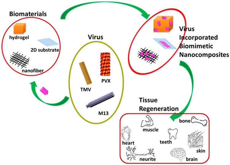
Owing to the astonishing properties of non-harmful viruses, tissue regeneration using virus-based biomimetic materials has been an emerging trend recently. The selective peptide expression and enrichment of the desired peptide on the surface, monodispersion, self-assembly, and ease of genetic and chemical modification properties have allowed viruses to take a long stride in biomedical applications. Researchers have published many reviews so far describing unusual properties of virus-based nanoparticles, phage display, modification, and possible biomedical applications, including biosensors, bioimaging, tissue regeneration, and drug delivery, however the integration of the virus into different biomaterials for the application of tissue regeneration is not yet discussed in detail. This review will focus on various morphologies of virus-incorporated biomimetic nanocomposites in tissue regeneration and highlight the progress, challenges, and future directions in this area.

Keywords: biomimetic nanocomposites; phage display; tissue regeneration; virus.


Conflict of interest statement

The authors declare no conflict of interest.

Figures

Figure 1
Figure 1
A compendium of tissue regeneration functionality of various virus incorporated biomimetic nanocomposites. Plant-based viruses Tobacco Mosaic Virus (TMV) and Potato Virus X (PVX) and bacteriophage M13 were shown. Reproduced with permission from [31]. Copyright Elsevier, 2010.
Figure 2
Figure 2
TEM micrographs of wild type Tobacco Mosaic Virus (TMV) nanoparticles, scale bar 200 nm (a) and TMV/PANI/PSS nanofiber (b) generated by flow assembly method, scale bar 500 nm. Reproduced with permission from [56]. Copyright American Chemical Society, 2015. (c) SEM micrograph of freeze-dried capsules of T4 bacteriophage/alginate water in oil emulsion in chloroform, scale bar 5 μm. (d) SEM and TEM micrographs (e) of PEO electrospun nanofiber containing T4 bacteriophage/alginate have been shown scale bars of 10 μm and 100 nm, respectively. The arrow indicates the presence of T4/alginate into the nanofiber. Reproduced with permission from [57]. Copyright John Wiley and Sons, 2013. (f) SEM image of the 3D printed bioceramic bone scaffold consisting of biphasic calcium phosphate with pores filled with a matrix of chitosan and RGD phage, scale bar 200 μm. Reproduced with permission from [58]. Copyright John Wiley and Sons, 2014. (g) SEM micrograph of porous alginate hydrogel containing TMV particles displaying interconnected channels and macropores, scale bar 100 μm. Reproduced with permission from [59]. Copyright American Chemical Society, 2012. (h) The TEM image of a mineralized E8-displaying phage bundle formed after 90 h incubation in the solution containing calcium and phosphate ions, scale bar 100 nm. Reproduced with permission from [60]. Copyright John Wiley and Sons, 2010. (i) The TEM image of the HAP-fd phage bundle with scale bar 100 nm. The circle indicates the presence of hydroxyapatite nanoparticles in the fibrous structure. Reproduced with permission from [61]. Copyright American Chemical Society, 2010.
Figure 3
Figure 3
(a) Field emission scanning electron micrographs of Baby Hamster Kidney cells after 1 h incubation on electrospun nanofibrous substrates PVA, PVA-TMV, and PVA-TMV/RGD. Scale bar 2 μm. Reproduced with permission from [71]. Copyright John Wiley and Sons, 2014. (b) Immunofluorescence staining of longitudinal sections of total cyst area in spinal cord injury (SCI)-treated female Sprague-Dawley rats during the chronic phase. The synthesis of GAP-43 immunopositive fibers was significantly greater in bone marrow homing peptide-expressed phages (4G-BMHP1) treated group when compared to control groups (SCI control and saline). Scale bar 400 mm. The percentage of immunopositive fibers into the cyst area per total cyst area is represented in a bar graph (c) from six independent experiment results, and the values are reported with ± SEM. Significant factor * p ˂ 0.05, 4G-BMHP1 vs. SCI control; 4G-BMHP1 vs. saline. Reproduced with permission from [72]. Copyright PLOS, 2011. (d) Bright-field optical microscope images of histochemical staining of alkaline phosphatase (ALPL) in bone marrow-derived stem cells (BMSCs) in TMV and phosphate grafted TMV (TMV-Phos)-coated Ti substrates after 14 days of culture. BMSCs have been shown to form neighboring well-spread cells under osteogenic conditions, which are stained highly positive for ALPL. Scale bar is 100 μm. (e,f) SEM micrographs and EDX analyses of TMV and TMV-Phos coating on Ti substrates. Scale bar for SEM is 100 μm. Reproduced with permission from [73]. Copyright Elsevier, 2010.

References

    1. Shafiee A., Atala A. Tissue engineering: Toward a new era of medicine. Ann. Rev. Med. 2017;68:29–40. doi: 10.1146/annurev-med-102715-092331. - DOI - PubMed
    1. Atala A. Regenerative medicine strategies. J. Pediatr. Surg. 2012;47:17–28. doi: 10.1016/j.jpedsurg.2011.10.013. - DOI - PubMed
    1. Raja I.S., Fathima N.N. Gelatin–cerium oxide nanocomposite for enhanced excisional wound healing. ACS Appl. Biol. Mater. 2018;1:487–495. doi: 10.1021/acsabm.8b00208. - DOI - PubMed
    1. Galliot B., Crescenzi M., Jacinto A. Trends in tissue repair and regeneration. Development. 2017;144:357–364. doi: 10.1242/dev.144279. - DOI - PubMed
    1. Dorati R., DeTrizio A., Modena T., Conti B., Benazzo F., Gastaldi G., Genta I. Biodegradable scaffolds for bone regeneration combined with drug-delivery systems in osteomyelitis therapy. Pharmaceuticals. 2017;10:96. doi: 10.3390/ph10040096. - DOI - PMC - PubMed

LinkOut - more resources