. 2019 Oct;11(7):1206-1218.
doi: 10.1080/19420862.2019.1632113. Epub 2019 Jul 16.

Selection and characterization of FcεRI phospho-ITAM specific antibodies

Affiliations

Selection and characterization of FcεRI phospho-ITAM specific antibodies

Nileena Velappan et al. MAbs. 2019 Oct.

Abstract

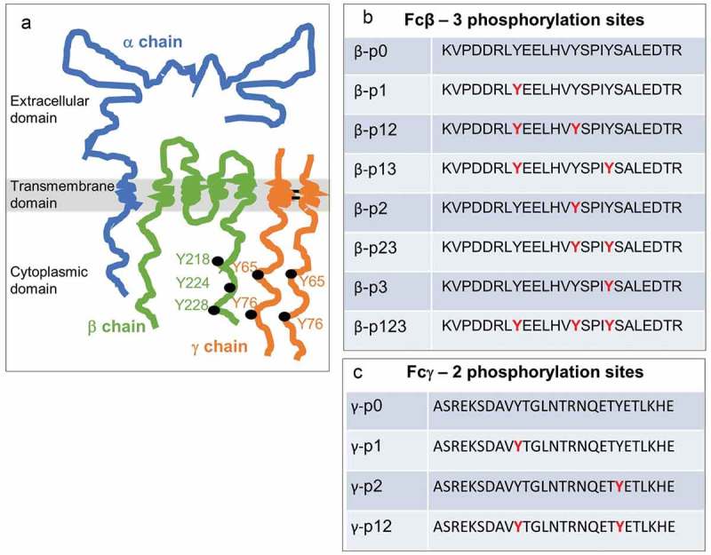
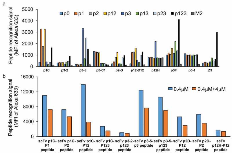
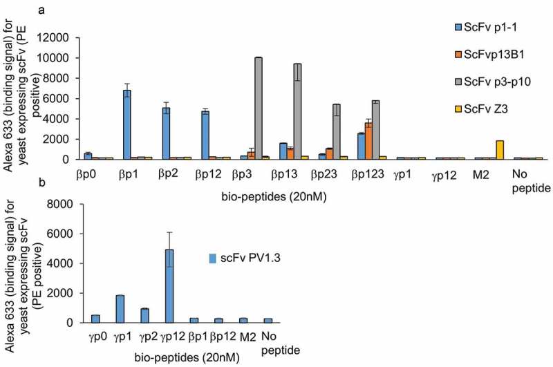
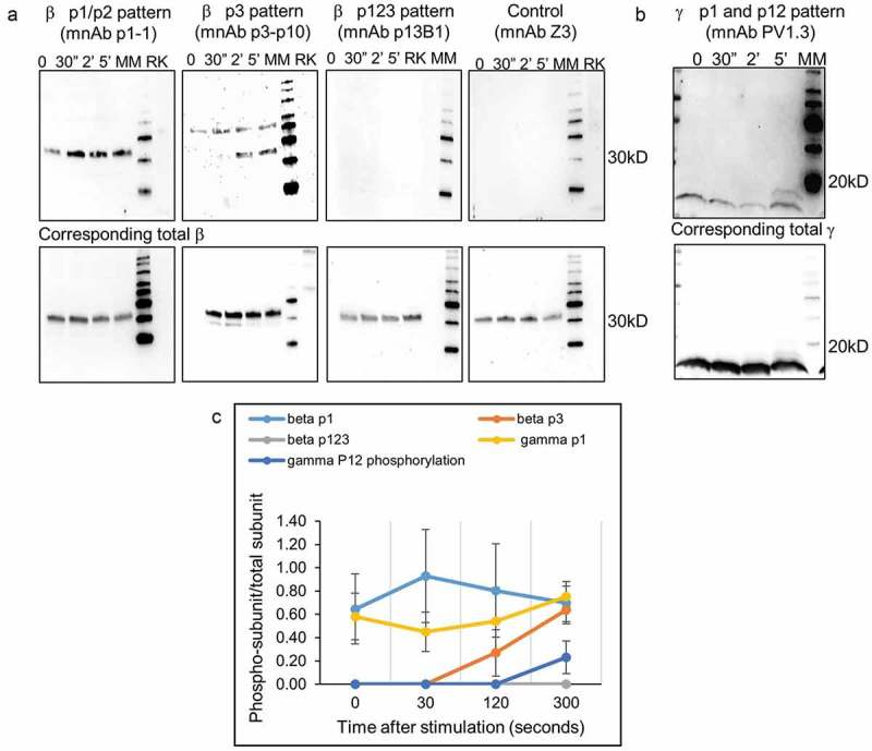
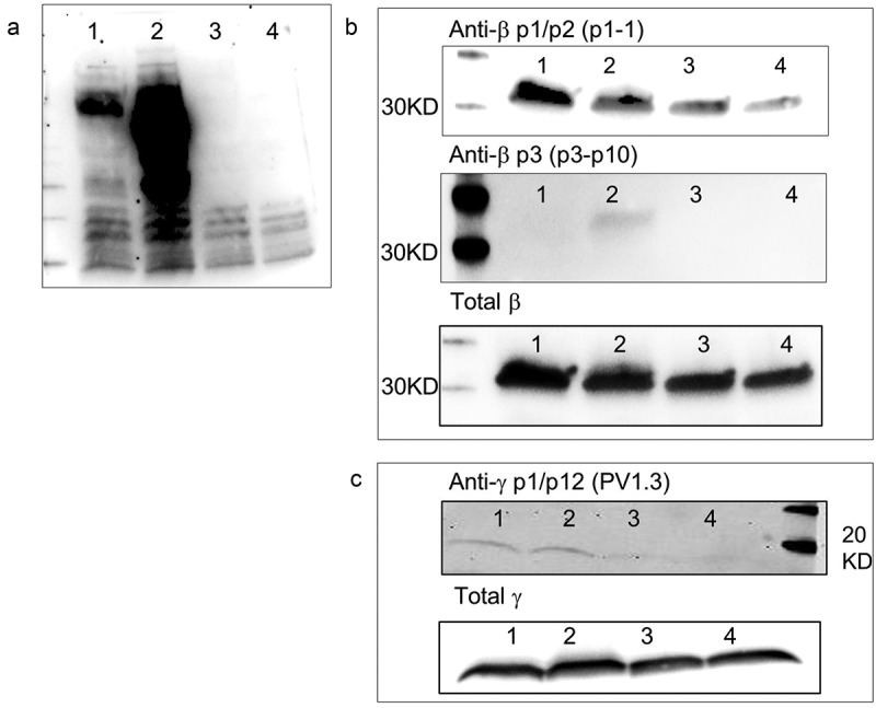
Post-translational modifications, such as the phosphorylation of tyrosines, are often the initiation step for intracellular signaling cascades. Pan-reactive antibodies against modified amino acids (e.g., anti-phosphotyrosine), which are often used to assay these changes, require isolation of the specific protein prior to analysis and do not identify the specific residue that has been modified (in the case that multiple amino acids have been modified). Phosphorylation state-specific antibodies (PSSAs) developed to recognize post-translational modifications within a specific amino acid sequence can be used to study the timeline of modifications during a signal cascade. We used the FcεRI receptor as a model system to develop and characterize high-affinity PSSAs using phage and yeast display technologies. We selected three β-subunit antibodies that recognized: 1) phosphorylation of tyrosines Y218 or Y224; 2) phosphorylation of the Y228 tyrosine; and 3) phosphorylation of all three tyrosines. We used these antibodies to study the receptor activation timeline of FcεR1 in rat basophilic leukemia cells (RBL-2H3) upon stimulation with DNP24-BSA. We also selected an antibody recognizing the N-terminal phosphorylation site of the γ-subunit (Y65) of the receptor and applied this antibody to evaluate receptor activation. Recognition patterns of these antibodies show different timelines for phosphorylation of tyrosines in both β and γ subunits. Our methodology provides a strategy to select antibodies specific to post-translational modifications and provides new reagents to study mast cell activation by the high-affinity IgE receptor, FcεRI.

Keywords: FcεR1 receptor subunits; Phosphorylation state-specific antibodies; phage display; post-translational modifications; yeast display.


Figures

Figure 1.
Figure 1.
FcϵR1 receptor subunits and corresponding ITAMs with associated phosphorylation patterns. a) Cartoon of the oligomeric (α,β,γ2) FcϵR1 complex. b-c) ITAMs in the β and γ subunits have three and two tyrosine phosphorylation sites (red), respectively. Panels b-c illustrate the set of combinations that can arise from partial or complete phosphorylation at these sites.
Figure 2.
Figure 2.
ScFv peptide and phosphate specificity assays upon completion of the initial selection process for the β ITAM peptides. a) Nine example scFvs obtained following three rounds of phage selection and three rounds of yeast sorting. Yeast displaying scFvZ3 and its antigen M2 peptide of Influenza A were used as negative controls in this flow-based assay. Binding assays were conducted at 100 nM peptide concentration. Data show varying levels of specificity of individual scFvs for different phospho-peptide(s). b) Five scFvs (p1C, p3-2, p3-5, p2D, and p12H) from panel A were evaluated for recognition of non-biotinylated (native) peptide using a competition assay. Paired plots show relative binding of biotinylated peptides to scFv displayed on yeast in the absence (blue) or presence (orange) of 10X non-biotinylated peptide.
Figure 3.
Figure 3.
Peptide and phosphate specificity assays for affinity-matured scFvs. a) Specificity assay for scFvs p1-1, p13B1, p3p10 against the eight β-phospho-peptides using yeast displayed scFvs in a flow-based assay. FcϵR1 γp1 and γp12 peptides and influenza M2 peptides were used as controls. The data also show binding profile for scFvZ3, which was used as negative control antibody. b) Specificity assay for anti-γ scFv PV1.3 against the four γ phospho-peptides with βp1, βp12, and M2 peptides used as controls.
Figure 4.
Figure 4.
Phosphorylation pattern analysis of FcϵR1 subunits. a) Assessment of phosphorylation patterns of β subunits after stimulation with DNP-BSA is shown using three antibodies p1-1, p13B1, and p3p10 utilizing western blotting with antibodies in minibody format. The FcϵR1 receptor was stimulated with 2 nM DNP-BSA and the data show phosphorylation patterns at various time points post stimulation. Phosphorylation at resting (basal) is also given. Antibody Z3 and rat kidney cell lysate (RK) were used as negative controls. The lower panel shows the total β detection used as a loading control. b) Similar analysis performed for anti-γ PV1.3. c) Patterns of phosphorylation graphed with data from multiple experiments (3–8 cell lysates prepared at different times).
Figure 5.
Figure 5.
Antibody recognition FcϵR1 subunits of untreated and phosphatase treated cell lysates. a) Presence of phospho-tyrosines in whole cell lysates at resting (#1), stimulated with DNP-BSA for 5 min (#2) and after these samples have been treated with tyrosine phosphatase for 30 min (#3 & 4), assayed using pan-reactive pY antibodies utilizing western blotting. b) Specificity of the p1-1 and p3p10 antibodies for phosphorylated β subunits were assessed by comparing recognition of pattern of cell lysates before and after phosphatase treatment. Total β subunit western analysis was performed as a loading control. c) Similar analysis performed for the anti-γ antibody PV1.3.

References

    1. Sibley DR, Benovic JL, Caron MG, Lefkowitz RJ.. Phosphorylation of cell surface receptors: A mechanism for regulating signal transduction pathway. Endocr Rev. 1988;9(1):38–56. doi:10.1210/edrv-9-1-38. - DOI - PubMed
    1. Cohen P. The origins of protein phosphorylation. Nat Cell Biol. 2002;4:E127–E130. doi:10.1038/ncb0502-e127. - DOI - PubMed
    1. Rabeh WM, Ribeiro A, Panovska J, Dessaud E, Sasai N, Page KM, Briscoe J, Ribes V. Correction of both NBD1 energetics and domain interface is required to restore ΔF508 CFTR folding and function. Cell. 2012;148:150–63. doi:10.1016/j.cell.2011.11.024. - DOI - PMC - PubMed
    1. Mandell JW. Phosphorylation state-specific antibodies: applications in investigative and diagnostic pathology. Am J Pathol. 2003;163:1687–98. doi:10.1016/S0002-9440(10)63525-0. - DOI - PMC - PubMed
    1. Mandell JW. Immunohistochemical assessment of protein phosphorylation state: the dream and the reality. Histochem Cell Biol. 2008;130:465–71. doi:10.1007/s00418-008-0474-z. - DOI - PMC - PubMed

Publication types

LinkOut - more resources