. 2019 Jul 24;286(1907):20191039.
doi: 10.1098/rspb.2019.1039. Epub 2019 Jul 17.

Convergent evolution of cytochrome P450s underlies independent origins of keto-carotenoid pigmentation in animals

Affiliations

Convergent evolution of cytochrome P450s underlies independent origins of keto-carotenoid pigmentation in animals

Nicky Wybouw et al. Proc Biol Sci. .

Abstract

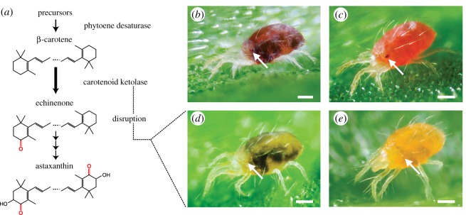
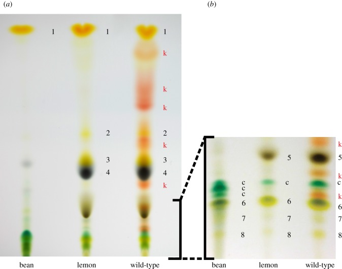
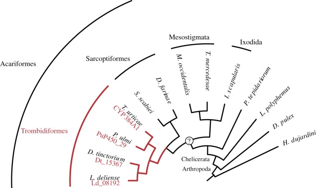
Keto-carotenoids contribute to many important traits in animals, including vision and coloration. In a great number of animal species, keto-carotenoids are endogenously produced from carotenoids by carotenoid ketolases. Despite the ubiquity and functional importance of keto-carotenoids in animals, the underlying genetic architectures of their production have remained enigmatic. The body and eye colorations of spider mites (Arthropoda: Chelicerata) are determined by β-carotene and keto-carotenoid derivatives. Here, we focus on a carotenoid pigment mutant of the spider mite Tetranychus kanzawai that, as shown by chromatography, lost the ability to produce keto-carotenoids. We employed bulked segregant analysis and linked the causal locus to a single narrow genomic interval. The causal mutation was fine-mapped to a minimal candidate region that held only one complete gene, the cytochrome P450 monooxygenase CYP384A1, of the CYP3 clan. Using a number of genomic approaches, we revealed that an inactivating deletion in the fourth exon of CYP384A1 caused the aberrant pigmentation. Phylogenetic analysis indicated that CYP384A1 is orthologous across mite species of the ancient Trombidiformes order where carotenoids typify eye and body coloration, suggesting a deeply conserved function of CYP384A1 as a carotenoid ketolase. Previously, CYP2J19, a cytochrome P450 of the CYP2 clan, has been identified as a carotenoid ketolase in birds and turtles. Our study shows that selection for endogenous production of keto-carotenoids led to convergent evolution, whereby cytochrome P450s were independently co-opted in vertebrate and invertebrate animal lineages.

Keywords: CYP384A1; carotenoid ketolase; convergent evolution; keto-carotenoids; lemon.


Conflict of interest statement

We declare we have no competing interests.

Figures

Figure 1.
Figure 1.
The conserved keto-carotenoid biosynthesis pathway in tetranychid mites and its disruption in lemon mutants. (a) The proposed pathway for carotenoid biosynthesis in spider mites [18] adapted to incorporate recent findings (endogenous synthesis of β-carotene by phytoene desaturase [4]). β-carotene is converted to echinenone, which leads to three major keto-carotenoids: 3-hydroxyechinenone and phoenicoxanthin (not shown), and astaxanthin [18]. Carotenoids are depicted in their de-esterified forms. (b) Wild-type T. kanzawai. (c) Wild-type diapausing T. kanzawai. (d) Lemon T. kanzawai. (e) Lemon diapausing T. kanzawai. Each panel depicts an adult female. Arrows highlight the anterior and posterior eye spots, which are red-coloured in wild-type individuals. The dark regions in feeding, non-diapausing mites are gut contents that are visible through the partially translucent cuticle. Feeding spots are absent (or nearly so) in diapausing mites that have ceased to actively feed. Scale bars represent 0.1 mm.
Figure 2.
Figure 2.
Lemon T. kanzawai lacks endogenously produced keto-carotenoids. Panels (a) and (b) show HPTLC plates for plant and mite extracts run with mobile phases of 20% and 25% acetone in hexane, respectively. Using previously determined Rf values and colour profiles [–18], we tentatively identified the carotenoid pigments as: 1: α- and β-carotene; k: keto-carotenoid (esterified in vivo); 2: β-carotene-diepoxide; 3: unknown epoxide; 4: chlorophyll a; 5: chlorophyll b; c: chlorophyll derivatives; 6: lutein and lutein 5,6-epoxide; 7: violaxanthin; 8: neoxanthin.
Figure 3.
Figure 3.
Lemon T. kanzawai accumulates higher levels of β-carotene. Panels (a) and (b) show the levels of β-carotene and astaxanthin, respectively, in wild-type and lemon T. kanzawai. Carotenoid levels were determined by HPLC for both feeding and diapausing adult female mites. N.D. stands for not detected. Error bars represent the standard errors, with a sample size of three.
Figure 4.
Figure 4.
Bulked segregant analysis locates the lemon locus and reveals a non-functional CYP384A1 as the genetic basis. (a) Differences in the frequencies of parental Jp-inbred-lemon alleles between each of the three lemon selected and one wild-type offspring pools are plotted in a sliding window analysis. The three T. urticae reference chromosomes are shown in alternating white and grey and are ordered by decreasing length. Dashed lines represent the 5% FDR for an association between parental Jp-inbred-lemon allele frequencies and the lemon phenotype. The maximal average allele frequency of the three replicates (i.e. the BSA peak) is located at cumulative genomic position 14 287 500. (b) CYP384A1 and a 3′ end fragment of its neighbouring gene reside in the minimal candidate region. Gene models and their genomic position are based on the T. urticae genome annotation, with exons and introns depicted as dark and light grey rectangles, respectively. Strands are represented as ‘+’ (forward) and ‘−’ (reverse). Blue triangles delineate the genomic position of the genetic markers used in the fine-mapping approach and the vertical dotted lines demarcate the 8.96 kb minimal candidate region. The green triangle highlights the location of the BSA peak. (c) Read coverage reveals a deletion within the CYP384A1 coding sequence in the three lemon selected offspring pools and parental Jp-inbred-lemon (black arrow). DNA sequence read coverage depth across the minimal candidate region is shown relative to the chromosome-wide average. (d) The deletion spans 246 bp within the fourth exon of the CYP384A1 coding sequence concomitant with 7 bp of inserted sequence. The five essential cytochrome P450 domains are plotted above the gene models.
Figure 5.
Figure 5.
CYP384A1 is orthologous across mite species of the Trombidiformes order. The maximum-likelihood phylogenetic reconstruction uncovered a 1 : 1 : 1 : 1 orthology of CYP384A1 for the four trombidiform mite species with available genomic resources (Arthropoda: Chelicerata: Acari: Acariformes: Trombidiformes) (electronic supplementary material, figure S5). The gene IDs for the identified orthologues are given in red font below the species name. A monophyletic origin for mites (Chelicerata: Acari) remains under debate [42].

References

    1. Goodwin TW. 1984. The biochemistry of the carotenoids, volume II: animals. London, UK: Chapman and Hall.
    1. Hill GE, McGraw KJ. 2006. Bird coloration, vol. 2: function and evolution. Cambridge, MA: Harvard University Press.
    1. Bosse TC, Veerman A. 1996. Involvement of vitamin A in the photoperiodic induction of diapause in the spider mite Tetranychus urticae is demonstrated by rearing an albino mutant on a semi-synthetic diet with and without p-carotene or vitamin A. Physiol. Entomol. 21, 188–192. (10.1111/j.1365-3032.1996.tb00854.x) - DOI
    1. Bryon A, et al. 2017. Disruption of a horizontally transferred phytoene desaturase abolishes carotenoid accumulation and diapause in Tetranychus urticae. Proc. Natl Acad. Sci. USA 114, E5871–E5880. (10.1073/pnas.1706865114) - DOI - PMC - PubMed
    1. Heath JJ, Cipollini DF, Stireman JO III. 2013. The role of carotenoids and their derivatives in mediating interactions between insects and their environment. Arthropod-Plant Interact. 7, 1–20. (10.1007/s11829-012-9239-7) - DOI

Publication types

MeSH terms