

Panniculitis-like T-cell-lymphoma in the mesentery associated with hemophagocytic syndrome: autopsy case report
- PMID: 31311562
- PMCID: PMC6636044
- DOI: 10.1186/s13000-019-0854-9
Panniculitis-like T-cell-lymphoma in the mesentery associated with hemophagocytic syndrome: autopsy case report
Abstract
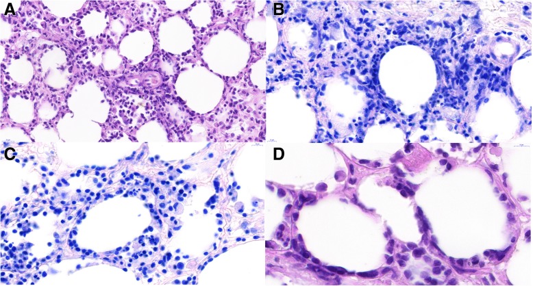
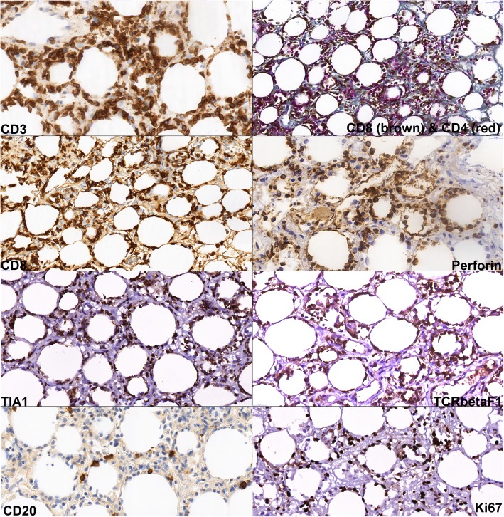
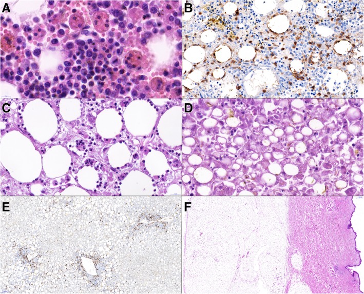
Background: Panniculitis-like T-cell lymphoma is an uncommon type of non-Hodgkin lymphoma, occurring usually in the form of nodules within the subcutaneous fat tissue of the extremities or trunk. In the literature, subcutaneous panniculitis-like T-cell lymphoma (SPTCL) is described as a distinct type of T-cell lymphoma with a variable clinical behavior, depending on molecular phenotype of T-cell receptor (TCR) and on the presence or absence of hemophagocytic syndrome.
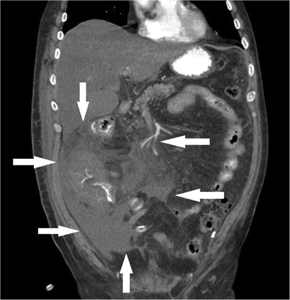
Case presentation: We present a bioptic and autoptic case of a 65-years old Caucasian man with panniculitic T-cell lymphoma with morphological and immunohistochemical features of SPTCL, limited to the retroperitoneal and mesenteric mass, i.e. without any cutaneous involvement, and associated with severe hemophagocytic lymphohistiocytosis.
Conclusion: A panniculitic T-cell lymphoma with morphological and molecular features of SPTCL, which is limited to mesentery, i.e. does not involve subcutaneous fat, seems to be exceedingly rare.
Keywords: Hemophagocytosis; Lymphohistiocytosis; Mesentery; Panniculitis; T-cell lymphoma.
Conflict of interest statement
The authors declare that they have no competing interests.
Figures
References
-
- Jaffe ES, Gaulard P, Cerroni L. Subcutaneous panniculitis-like T-cell lymphoma. In: Swerdlow SH, Campo E, Harris NL, Jaffe ES, Pileri SA, Stein H, Thiele J, editors. WHO Classification of Tumours of Haematopoetic and Lymphoid Tissues (Revised 4th edition) Lyon: IARC; 2017. pp. 383–385.
-
- Willemze R, Jansen PM, Cerroni L, Berti E, Santucci M, Assaf C, et al. Subcutaneous panniculitis-like T-cell lymphoma: definition, classification, and prognostic factors: an EORTC cutaneous lymphoma group study of 83 cases. Blood. 2008;111(2):838–845. doi: 10.1182/blood-2007-04-087288. - DOI - PubMed
-
- Salhany KE, Macon WR, Choi JK, Elenitsas R, Lessin SR, Felgar RE, et al. Subcutaneous panniculitis-like T-cell lymphoma: clinicopathologic, immunophenotypic, and genotypic analysis of alpha/beta and gamma/delta subtypes. Am J Surg Pathol. 1998;22(7):881–893. doi: 10.1097/00000478-199807000-00010. - DOI - PubMed
Publication types
MeSH terms
Supplementary concepts
Grants and funding
LinkOut - more resources
Full Text Sources
Research Materials
