. 2019 Jul 16;38(1):311.
doi: 10.1186/s13046-019-1293-x.

G-quadruplex ligand RHPS4 radiosensitizes glioblastoma xenograft in vivo through a differential targeting of bulky differentiated- and stem-cancer cells

Affiliations

G-quadruplex ligand RHPS4 radiosensitizes glioblastoma xenograft in vivo through a differential targeting of bulky differentiated- and stem-cancer cells

F Berardinelli et al. J Exp Clin Cancer Res. .

Abstract

Background: Glioblastoma is the most aggressive and most lethal primary brain tumor in the adulthood. Current standard therapies are not curative and novel therapeutic options are urgently required. Present knowledge suggests that the continued glioblastoma growth and recurrence is determined by glioblastoma stem-like cells (GSCs), which display self-renewal, tumorigenic potential, and increased radio- and chemo-resistance. The G-quadruplex ligand RHPS4 displays in vitro radiosensitizing effect in GBM radioresistant cells through the targeting and dysfunctionalization of telomeres but RHPS4 and Ionizing Radiation (IR) combined treatment efficacy in vivo has not been explored so far.

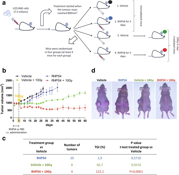
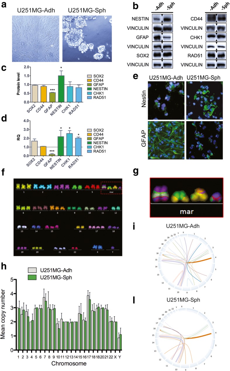
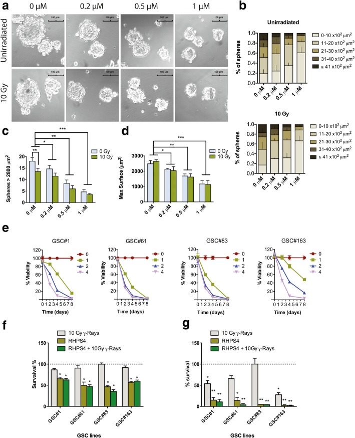
Methods: RHPS4 and IR combined effects were tested in vivo in a heterotopic mice xenograft model and in vitro in stem-like cells derived from U251MG and from four GBM patients. Cell growth assays, cytogenetic analysis, immunoblotting, gene expression and cytofluorimetric analysis were performed in order to characterize the response of differentiated and stem-like cells to RHPS4 and IR in single and combined treatments.

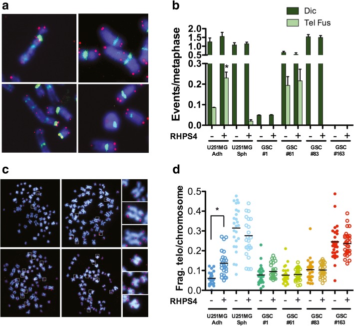
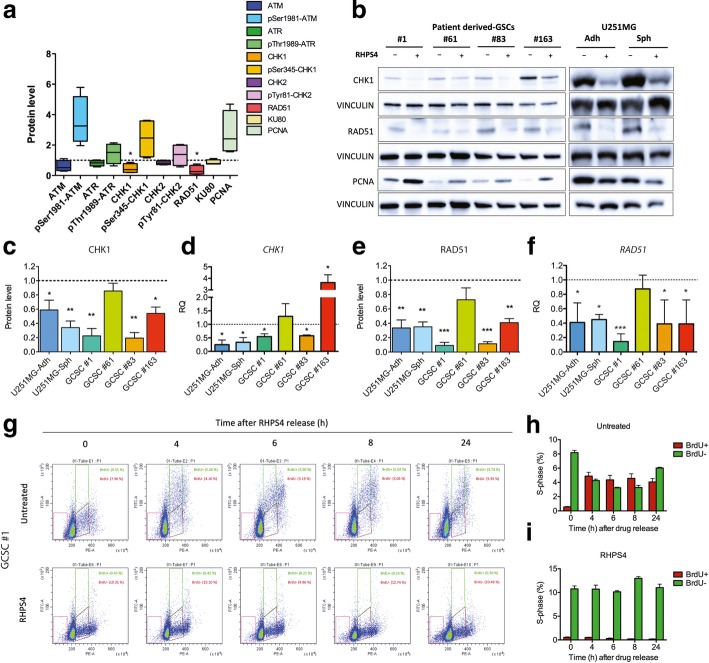
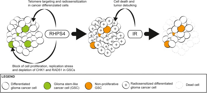
Results: RHPS4 administration and IR exposure is very effective in blocking tumor growth in vivo up to 65 days. The tumor volume reduction and the long-term tumor control suggested the targeting of the stem cell compartment. Interestingly, RHPS4 treatment was able to strongly reduce cell proliferation in GSCs but, unexpectedly, did not synergize with IR. Lack of radiosensitization was supported by the GSCs telomeric-resistance observed as the total absence of telomere-involving chromosomal aberrations. Remarkably, RHPS4 treatment determined a strong reduction of CHK1 and RAD51 proteins and transcript levels suggesting that the inhibition of GSCs growth is determined by the impairment of the replication stress (RS) response and DNA repair.

Conclusions: We propose that the potent antiproliferative effect of RHPS4 in GSCs is not determined by telomeric dysfunction but is achieved by the induction of RS and by the concomitant depletion of CHK1 and RAD51, leading to DNA damage and cell death. These data open to novel therapeutic options for the targeting of GSCs, indicating that the combined inhibition of cell-cycle checkpoints and DNA repair proteins provides the most effective means to overcome resistance of GSC to genotoxic insults.

Keywords: G4 ligands; Glioma stem-like cells; RHPS4; Radiosensitization; Telomeres.


Conflict of interest statement

The authors declare that they have no competing interests.

Figures

Fig. 1
Fig. 1
RHPS4 and IR synergize to inhibit the growth of glioblastoma xenograft preventing tumor relapse. Graphical representation of the in vivo experimental plan. U251MG cell xenografted mice were randomized in four groups: Vehicle, RHPS4, Vehicle + 10 Gy and RHPS4 + 10 Gy. RHPS4 (10 mg/kg per day) or PBS (vehicle) were administered through intravenous injection for 5 days, then mice were irradiated with a single dose of 10 Gy of X-rays (a). The graph in b shows the tumor growth kinetic relative to each treated group started when tumor mass reached 800 mm3 in volume. In panel c is shown the tumor growth inhibition (TGI%) of treated tumors for each experimental group compared with vehicle group. Representative images of U251MG cell xenografted mice 65 days post-treatment with a clear regression of tumor mass in the combined treatment group (RHPS4 + 10 Gy) (d)
Fig. 2
Fig. 2
Stem cell markers and cytogenetic characterization of U251MG-Sph. Representative images of adherent U251MG cells and spheres derived from the same cell line (a). Western blot of NESTIN, SOX2, CD44 and GFAP in U251MG-Ahd and -Sph cells (b). Densitometric analysis revealed a significant reduction of GFAP and a significant increase of NESTIN in U251MG-Sph compared to U251MG-Adh (c). Data were also confirmed by qRT-PCR (d). Images of immunofluorescence versus NESTIN and GFAP confirmed western blot data (e). Most common karyogram observed in U251MG-Adh cells as revealed by mFISH (f). Derivative chromosomes are indicated as mar and involved chromosomes 11–10-15, 10–15, 16–4 and 16–3 (g). Ploidy of U251MG-Adh and -Sph was completely superimposable (h), whereas as shown in circos graphs, frequency of chromosomal exchanges is higher in U251MG-Adh (i) than in U251MG-Sph (l). * P < 0.05, ** P < 0.01, *** P < 0.001 (Student’s t-test)
Fig. 3
Fig. 3
Cytotoxic effect of RHPS4 in single treatment and in combination with IR. Images of U251MG-derived neurospheres treated with increasing concentrations of RHPS4 (0.2–1 μM for 5 days) and then exposed to 10 Gy of X-rays (a). Neurospheres maximal surface was automatically calculated by IS-Capture software after manual surrounding of each sphere. Maximal surface data were grouped in 5 different classes (0–10, 11–20, 21–30, 31–40 and ≥ 41*102 μm2) for both unirradiated and 10 Gy-exposed U251MH-Sph cells. Data have been reported as percentage of the total number of spheres analyzed and represent mean values ± s.d. (n = 3) (b). Spheres number and max surface in samples exposed to RHPS4 and IR were shown in (c) and (d), respectively. Data represent mean values ± s.d. (n = 3). Growth curves of GSCs from patients treated with RHPS4 (1, 2, 3 and 4 μM) and followed for 8 days (e). Data represent mean values ± s.d. (n = 2). Effect of RHPS4 and γ-rays combined treatment on cell growth in GSCs from patients. Cell growth was evaluated after 3 (f) and 7 days (g) from irradiation. Combined treatment was performed treating cells with IC25 calculated at 4 days and then exposing them to 10 Gy γ-rays. Data represent mean values ± s.d. (n = 2). * P < 0.05, ** P < 0.01, *** P < 0.001 (Student’s t-test)
Fig. 4
Fig. 4
RHPS4 induces telomeric fusions and telomeric fragility in glioblastoma differentiated cells but not in GSCs. Representative images of telomere fusions involved in the formation of dicentric, tricentric and ring chromosomes observed in the U251MG-Adh cell line treated with 0.5 μM RHPS4 for 5 days (a). Frequency of classical dicentrics (dic) and dicentrics generated from telomere fusions (tel fus) in the U251MG derived cell lines and in GSCs lines from patients (b). Data represent mean values ± s.d. (n = 2). Representative images of U251MG-Adh cells in which are present several fragile telomeres (surrounded by boxes). Some of them were enlarged on the right side of the Fig. (c). Frequency of fragile telomeres per chromosome in the U251MG derived cell lines and in GSCs lines obtained from patients treated or not with RHPS4. Data represent the frequency of each metaphase scored and black bars denotes mean values (d). (n = 2). * P < 0.05 (Student’s t-test)
Fig. 5
Fig. 5
RHPS4 induces reduction of CHK1 and RAD51 and determine S-phase blockage in GSCs. Analysis of proteins involved in DNA damage response and checkpoint activation in patient derived-GSCs (a). Representative blots for CHK1, RAD51 and PCNA in patient derived-GSCs and U251MG (−Adh and -Sph) (b). CHK1 and RAD51 protein level and gene expression in all the cell lines analyzed (c, d, e, f). Data represent mean ± s.d. (n = 3). * P < 0.05, ** P < 0.01, *** P < 0.001 (Student’s t-test). Assessment of the BrdU-incorporating cells in samples exposed for 4 days to RHPS4. Cells were pulsed for 3 h with BrdU and, after RHPS4 release, cells were chased for additional 24 h (cell were fixed after 4, 6, 8 and 24 h) (g). Quantification of BrdU positive (BrdU+) and negative (BrdU-) cells in untreated (h) and RHPS4-reated cells (i). Note the total depletion of BrdU+ observed over-time in RHPS4 treated samples
Fig. 6
Fig. 6
RHPS4 inhibits glioblastoma cell proliferation through a differential targeting of bulky differentiated- and cancer stem-like cells. RHPS4 differently targets differentiated and GSCs cells. Differentiated glioblastoma cells (white circles) are radiosensitized (white circles marked with plus symbol) through the induction of telomere damage and dysfunctionalization [16, 17]. On the other hand, in GSCs (green circles) RHPS4 strongly reduces CHK1 and RAD51 protein levels leading to S-phase blockage, inhibition of cell proliferation (orange circles) and very likely to cell death. The subsequent exposure to IR determines the cell killing of differentiated (radiosensitized) cells and contributes to the tumor debulking. This model fits very well with data obtained in vivo, indicating that RHPS4 and IR combined treatment avoids tumor relapse and reduce tumor mass also in case of full blown and well-rooted tumors

Similar articles

Cited by

References

    1. Miller CR, Glioblastoma PA. Morphologic and molecular genetic diversity. Arch Pathol Lab Med. 2007;131:397–406. - PubMed
    1. Smoll NR, Schaller K, Gautschi OP. Long-term survival of patients with glioblastoma multiforme (GBM) J Clin Neurosci Elsevier Ltd. 2013;20:670–675. doi: 10.1016/j.jocn.2012.05.040. - DOI - PubMed
    1. Stupp R, Mason WP, Van Den Bent MJ, Weller M, Fisher B, Taphoorn MJB, et al. Radiotherapy plus concomitant and adjuvant Temozolomide for Glioblastoma. N Engl J Med. 2005;352:987–996. doi: 10.1056/NEJMoa043330. - DOI - PubMed
    1. Field KM, Drummond KJ, Yilmaz M, Tacey M, Compston D, Gibbs P, et al. Clinical trial participation and outcome for patients with glioblastoma: multivariate analysis from a comprehensive dataset. J Clin Neurosci. Elsevier Ltd. 2013;20:783–789. doi: 10.1016/j.jocn.2012.09.013. - DOI - PubMed
    1. Singh SK, Clarke ID, Terasaki M, Bonn VE, Hawkins C, Squire J, et al. Identification of a cancer stem cell in human brain tumors. Cancer Res. 2003;63:5821–5828. - PubMed

MeSH terms