

Compressive mechanical properties of rat and pig optic nerve head
- PMID: 31311622
- PMCID: PMC6698410
- DOI: 10.1016/j.jbiomech.2019.06.014
Compressive mechanical properties of rat and pig optic nerve head
Abstract
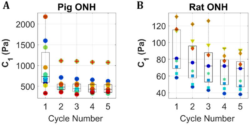
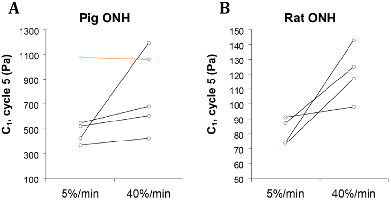
Glaucoma is the leading cause of irreversible blindness worldwide. Elevated intraocular pressure (IOP), the primary risk factor for glaucoma, is thought to induce abnormally high strains in optic nerve head (ONH) tissues, which ultimately result in retinal ganglion cell damage and vision loss. The mechanisms by which excessive deformations result in vision loss remain incompletely understood. The ability of computational and in vitro models of the ONH to provide insight into these mechanisms, in many cases, depends on our ability to replicate the physiological environment, which in turn requires knowledge of tissue biomechanical properties. The majority of mechanical data published to date regarding the ONH has been obtained from tensile testing, yet compression has been shown to be the main mode of deformation in the ONH under elevated IOP. We have thus tested pig and rat ONH tissue using unconfined cyclic compression. The material constants C1, obtained from fitting the stress vs. strain data with a neo-Hookean material model, were 428 [367, 488] Pa and 64 [53, 76] Pa (mean [95% Confidence Interval]) for pig and rat optic nerve head, respectively. Additionally, we investigated the effects of strain rate and tissue storage on C1 values. These data will inform future efforts to understand and replicate the in vivo biomechanical environment of the ONH.
Keywords: Compression; Mechanical properties; Neo-hookean; Optic nerve head.
Copyright © 2019 Elsevier Ltd. All rights reserved.
Conflict of interest statement
Conflict of Interest
The authors confirm that there are no conflicts of interest.
Figures

References
-
- Braunsmann C, Hammer CM, Rheinlaender J, Kruse FE, Schaffer TE, Schlotzer-Schrehardt U, 2012. Evaluation of lamina cribrosa and peripapillary sclera stiffness in pseudoexfoliation and normal eyes by atomic force microscopy. Invest Ophthalmol Vis Sci 53, 2960–2967. - PubMed
-
- Burgoyne CF, Downs JC, Bellezza AJ, Suh JK, Hart RT, 2005. The optic nerve head as a biomechanical structure: a new paradigm for understanding the role of IOP-related stress and strain in the pathophysiology of glaucomatous optic nerve head damage. Prog Retin Eye Res 24, 39–73. - PubMed
-
- Edwards ME, Good TA, 2001. Use of a mathematical model to estimate stress and strain during elevated pressure induced lamina cribrosa deformation. Curr Eye Res 23, 215–225. - PubMed
MeSH terms
Grants and funding
LinkOut - more resources
Full Text Sources
