. 2019 Jul 30;116(31):15453-15462.
doi: 10.1073/pnas.1903949116. Epub 2019 Jul 16.

A real-time, click chemistry imaging approach reveals stimulus-specific subcellular locations of phospholipase D activity

Affiliations

A real-time, click chemistry imaging approach reveals stimulus-specific subcellular locations of phospholipase D activity

Dongjun Liang et al. Proc Natl Acad Sci U S A. .

Abstract

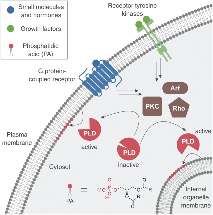
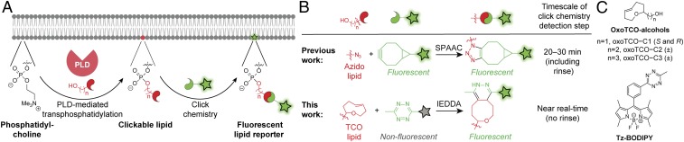
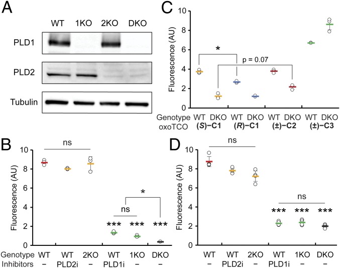
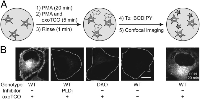
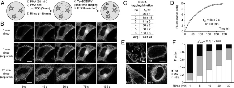
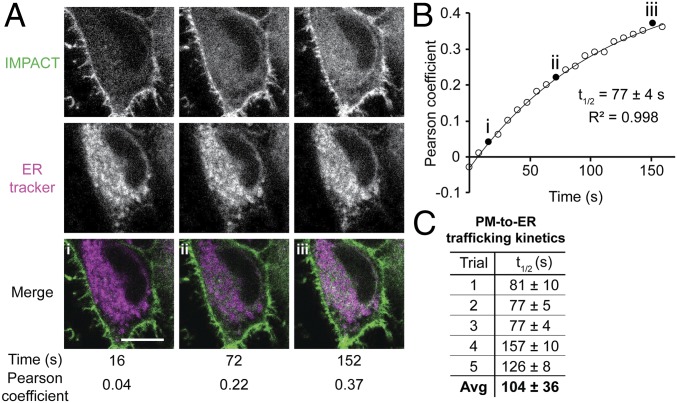
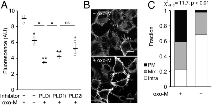
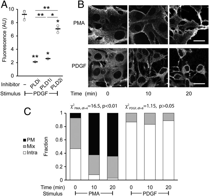
The fidelity of signal transduction requires spatiotemporal control of the production of signaling agents. Phosphatidic acid (PA) is a pleiotropic lipid second messenger whose modes of action differ based on upstream stimulus, biosynthetic source, and site of production. How cells regulate the local production of PA to effect diverse signaling outcomes remains elusive. Unlike other second messengers, sites of PA biosynthesis cannot be accurately visualized with subcellular precision. Here, we describe a rapid, chemoenzymatic approach for imaging physiological PA production by phospholipase D (PLD) enzymes. Our method capitalizes on the remarkable discovery that bulky, hydrophilic trans-cyclooctene-containing primary alcohols can supplant water as the nucleophile in the PLD active site in a transphosphatidylation reaction of PLD's lipid substrate, phosphatidylcholine. The resultant trans-cyclooctene-containing lipids are tagged with a fluorogenic tetrazine reagent via a no-rinse, inverse electron-demand Diels-Alder (IEDDA) reaction, enabling their immediate visualization by confocal microscopy in real time. Strikingly, the fluorescent reporter lipids initially produced at the plasma membrane (PM) induced by phorbol ester stimulation of PLD were rapidly internalized via apparent nonvesicular pathways rather than endocytosis, suggesting applications of this activity-based imaging toolset for probing mechanisms of intracellular phospholipid transport. By instead focusing on the initial 10 s of the IEDDA reaction, we precisely pinpointed the subcellular locations of endogenous PLD activity as elicited by physiological agonists of G protein-coupled receptor and receptor tyrosine kinase signaling. These tools hold promise to shed light on both lipid trafficking pathways and physiological and pathological effects of localized PLD signaling.

Keywords: click chemistry; lipid trafficking; phosphatidic acid; phospholipase D; second messengers.


Conflict of interest statement

The authors declare no conflict of interest.

Figures

Fig. 1.
Fig. 1.
Phospholipase D (PLD) enzymes are stimulated by different classes of cell-surface receptors through intracellular effectors, including those from the protein kinase C (PKC), Rho GTPase, and Arf GTPase families, to catalyze phosphatidic acid (PA) formation at different intracellular membrane locations. R and R′ denote the fatty acyl tails.
Fig. 2.
Fig. 2.
Overview of the imaging phospholipase D (PLD) activity with clickable alcohols via transphosphatidylation (IMPACT) method for visualizing PLD activity in live cells. (A) Cartoon schematic of IMPACT. (B) Comparison of established IMPACT method using azido alcohols and strain-promoted azide–alkyne cycloaddition (SPAAC) detection with the advance reported here, real-time (RT) IMPACT, using trans-cyclooctene (TCO)-containing alcohols and inverse electron-demand Diels–Alder (IEDDA) detection using tetrazine (Tz) reagents. (C) Structures of trans-5-oxocene (oxoTCO) alcohols and the fluorogenic Tz–BODIPY conjugate used in this study.
Fig. 3.
Fig. 3.
oxoTCO alcohols are effective and selective reporters of endogenous human PLD activity. (A and B) Generation and validation of PLD1 knockout (1KO), PLD2 knockout (2KO), or PLD1/2 double knockout (DKO) HeLa cells made by using CRISPR/Cas9-mediated mutagenesis. (A) Knockout was verified by Western blot analysis for PLD1, PLD2, or as a loading control, α-tubulin. (B) The indicated cells were pretreated with the indicated PLD inhibitor (PLD1i, VU0359595; or PLD2i, VU0364739) or DMSO for 30 min, followed by transphosphatidylation with 3-azido-1-propanol (1 mM) under PMA stimulation (100 nM) for 20 min, rinsing with PBS solution, and labeling with a cyclooctyne–BODIPY conjugate (1 µM) for 10 min, rinsing for 20 min, and analysis by flow cytometry. Indicated are mean fluorescence intensities in arbitrary units (AU). (C and D) Evaluation of oxoTCO alcohols as selective probes for endogenous PLD enzymes. Shown are mean fluorescence intensities from flow cytometry experiments wherein IMPACT labeling was performed as in A, except using the indicated oxoTCO alcohol (C) or (S)-oxoTCO–C1 (3 mM, 5 min; D) in place of 3-azido-1-propanol and Tz–BODIPY (0.33 µM, 1 min, no subsequent rinse) in place of cyclooctyne–BODIPY. Statistical significance was assessed by using a 1-way ANOVA followed by Games–Howell post hoc analysis. Asterisks directly above data points denote statistical significance compared with WT without the presence of inhibitors, asterisks above horizontal lines denote statistical significance comparing the 2 indicated data groups, and error bars represent SD (*P < 0.05 and ***P < 0.001; ns, not significant).
Fig. 4.
Fig. 4.
IMPACT with (S)-oxoTCO–C1 reveals the PM localization of active PLD enzymes stimulated with a phorbol ester. (A) Experimental setup: endogenous PLD enzymes were stimulated with PMA (100 nM, 20 min), followed by transphosphatidylation with (S)-oxoTCO–C1 (3 mM, 5 min, in the presence of PMA), rinse (1 min), and IEDDA reaction with Tz–BODIPY (0.33 µM, 1 min), followed by confocal microscopy imaging. (B) Representative images of HeLa cells labeled as described in A with the indicated negative controls. Where indicated, PLDi (the pan-PLD inhibitor FIPI) was applied 30 min before and during the transphosphatidylation step. DKO, PLD1/2 double knockout cells. Far right: cells were rinsed after the IEDDA reaction for 20 min before imaging. White dotted lines indicate cell outlines in the negative controls. (Scale bar: 10 μm.)
Fig. 5.
Fig. 5.
Real-time IMPACT enables precise determination of the subcellular localization of active PLD enzymes. (A) The experimental setup for RT-IMPACT is the same as indicated in Fig. 4 except that IEDDA reaction with Tz–BODIPY is monitored in real time by time-lapse confocal microscopy. (B) Representative images from time-lapse RT-IMPACT imaging of HeLa cells. The rinse (chase step) following the transphosphatidylation with (S)-oxoTCO–C1 and before IEDDA tagging was 1 min (Top and Middle) or 20 min (Bottom). (Top) Images are shown with identical intensity settings to facilitate visualization of reaction progress. (Middle and Bottom) Fluorescence intensities of the 9-, 15-, and 30-s time points have been brightened to facilitate visual comparison of localization of the IMPACT signal at different time points. (C and D) Quantification of the kinetics of the IEDDA tagging reaction based on total cellular fluorescence from 6 independent experiments (where the graph represents trial 5, with circles indicating data points, the curve indicating an exponential fit, and R2 indicating the coefficient of determination of the fit) with n = 8 to 16 cells for each trial and average (Avg) reaction half-life (t1/2) indicated as mean ± SD. (E and F) Quantification of the subcellular localizations of RT-IMPACT fluorescent signal at the 9-s time point of the IEDDA reaction as a function of the posttransphosphatidylation chase step, i.e., the rinse time after (S)-oxoTCO–C1 labeling and before IEDDA tagging. Cells were blindly scored as having plasma membrane (PM, black), intracellular (Intra, white), or a combination of PM and intracellular (Mix, gray) IMPACT fluorescence. y axis indicates fraction of cells with the indicated localization. (E, Top Left) Representative zoomed-out image of a population of cells at the 9-s time point from a 20-min rinse time point from F, with examples of PM (i), mix (ii), and intra (iii) localizations indicated in the other panels. (Scale bars: B and E, iiii, 10 µm; E, Top Left, 20 μm.) Statistical significance was assessed by using a χ2 test for independence, with the χ2 value (df, degrees of freedom) and associated P value indicated. Each bar contains data of 3 to 5 biological replicates with n = 30 to 72 total cells.
Fig. 6.
Fig. 6.
Real-time IMPACT reveals PM-to-ER trafficking of fluorescent lipids. (A) Representative images of HeLa cells from a 2-color time-lapse movie. HeLa cells were incubated with ER tracker, PMA, and (S)-oxoTCO–C1 for 5 min and rinsed for 1 min before time-lapse imaging. Values of Pearson correlation coefficients of colocalization between IMPACT (green) and ER tracker (magenta) were determined. Colocalization appears as white in merge. (B) Representative exponential fitting of kinetics of PM-to-ER trafficking. Plotted is increase in colocalization (Pearson coefficient) between IMPACT and ER tracker over time. Circles are data points, and the curve indicates an exponential fit. Filled circles (iiii) correspond to Pearson coefficients for the images shown in A, and R2 indicates the coefficient of determination of the fit. (C) Summary of quantification of PM-to-ER trafficking kinetics from 5 independent experiments (where the graph in B represents trial 3) with n = 8 to 16 cells for each trial and average (Avg) half-life of internalization (t1/2) indicated as mean ± SD. (Scale bar: 10 µm.)
Fig. 7.
Fig. 7.
Muscarinic M1 receptor (M1R) activation leads to PLD activity that is predominantly at the plasma membrane. HeLa cells stably expressing M1R were labeled for RT-IMPACT by treatment with the indicated PLD inhibitor or DMSO for 30 min (A only) and then simultaneous treatment of (S)-oxoTCO–C1 in the presence or absence of oxo-M (5 min), rinsing (1 min), and IEDDA reaction with Tz–BODIPY (0.33 µM) for 1 min followed by flow cytometry analysis (A), with mean fluorescence intensity in arbitrary units (AU) indicated, or (B and C) in real time with time-lapse confocal images taken (B) and quantified (C) 9 s after the addition of Tz–BODIPY, using the PM/Mix/Intra rubric described in Fig. 5. In B, the brightness of the –oxo-M image was increased to facilitate comparison of the localization of IMPACT-derived fluorescence in the 2 images. (Scale bars: 20 µm.) For A, statistical significance was assessed by using 1-way ANOVA followed by Games–Howell post hoc analysis. Asterisks directly above data points (*P < 0.05 and **P < 0.01; ns, not significant) denote statistical significance compared with the first sample (−inhibitor, +oxo-M), and error bars represent SD. For C, statistical significance was assessed by using a χ2 test for independence, with the χ2 value (df, degrees of freedom) and associated P value indicated. Each bar contains data of 3 (−oxo-M) or 8 (+oxo-M) biological replicates, with n = 41 (−oxo-M) or 112 (+oxo-M) total cells.
Fig. 8.
Fig. 8.
Platelet-derived growth factor (PDGF) receptor activation leads to intracellular PLD activity. NIH 3T3 cells were labeled for RT-IMPACT by treatment with the indicated PLD inhibitor or DMSO for 30 min (A only). Cells were stimulated with PDGF or PMA for the indicated time (0 to 20 min) followed by addition of (S)-oxoTCO–C1 in the continued presence of PDGF or PMA (5 min) and then rinsed (1 min), and the IEDDA reaction with Tz–BODIPY (0.33 µM) was performed for 1 min followed by flow cytometry analysis (A), with mean fluorescence intensity in arbitrary units (AU) indicated, or (B and C) in real time with time-lapse confocal images taken (B) and quantified (C) 9 s after the addition of Tz–BODIPY, using the PM/Mix/Intra rubric described in Fig. 5. (Scale bars: 20 µm.) For A, statistical significance was assessed by using 1-way ANOVA followed by Games–Howell post hoc analysis. Asterisks directly above data points (*P < 0.05 and **P < 0.01) denote statistical significance compared with the first sample (−inhibitor), asterisks above horizontal lines denote statistical significance comparing the 2 indicated samples, and error bars represent SD. For C, statistical significance was assessed by using a χ2 test for independence, with the χ2 value (df, degrees of freedom) and associated P value indicated. Each bar contains data of 3 to 4 biological replicates with n = 58 to 78 total cells for PDGF and 3 to 6 biological replicates, with n = 41 to 95 cells for PMA.

References

    1. Giepmans B. N. G., Adams S. R., Ellisman M. H., Tsien R. Y., The fluorescent toolbox for assessing protein location and function. Science 312, 217–224 (2006). - PubMed
    1. Oldach L., Zhang J., Genetically encoded fluorescent biosensors for live-cell visualization of protein phosphorylation. Chem. Biol. 21, 186–197 (2014). - PMC - PubMed
    1. Chakraborty K., Veetil A. T., Jaffrey S. R., Krishnan Y., Nucleic acid-based nanodevices in biological imaging. Annu. Rev. Biochem. 85, 349–373 (2016). - PMC - PubMed
    1. Chan J., Dodani S. C., Chang C. J., Reaction-based small-molecule fluorescent probes for chemoselective bioimaging. Nat. Chem. 4, 973–984 (2012). - PMC - PubMed
    1. Liu Y., Su Y., Wang X., “Phosphatidic acid-mediated signaling” in Lipid-Mediated Protein Signaling (Springer Netherlands, Dordrecht, 2013), pp. 159–176. - PubMed

Publication types

LinkOut - more resources