

Comparison of the Diagnostic Ability of Endoscopic Ultrasonography and Abdominopelvic Computed Tomography in the Diagnosis of Gastric Subepithelial Tumors
- PMID: 31311912
- PMCID: PMC6900302
- DOI: 10.5946/ce.2019.019
Comparison of the Diagnostic Ability of Endoscopic Ultrasonography and Abdominopelvic Computed Tomography in the Diagnosis of Gastric Subepithelial Tumors
Abstract
Background/aims: Endoscopic ultrasonography (EUS) is the most efficient imaging modality for gastric subepithelial tumors (SETs). However, abdominopelvic computed tomography (APCT) has other advantages in evaluating the characteristics, local extension, or invasion of SETs to adjacent organs. This study aimed to compare the diagnostic ability of EUS and APCT based on surgical histopathology results.
Methods: We retrospectively reviewed data from 53 patients who underwent both EUS and APCT before laparoscopic wedge resection for gastric SETs from January 2010 to December 2017 at a single institution. On the basis of histopathology results, we assessed the diagnostic ability of the 2 tests.
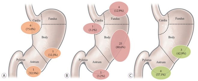
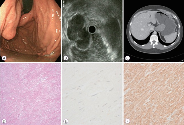
Results: The overall accuracy of EUS and APCT was 64.2% and 50.9%, respectively. In particular, the accuracy of EUS vs. APCT for the diagnosis of gastrointestinal stromal tumors (GISTs), leiomyomas, and ectopic pancreas was 83.9% vs. 74.2%, 37.5% vs. 0.0%, and 57.1% vs. 14.3%, respectively. Most of the incorrect diagnoses with EUS involved hypoechoic lesions originating in the fourth echolayer, with the most common misdiagnosed lesions being GISTs mistaken for leiomyomas and vice versa.
Conclusion: APCT showed a lower overall accuracy than EUS; however, APCT remains a useful modality for malignant/potentially malignant gastric SETs.
Keywords: Abdominopelvic computed tomography; Diagnosis; Endoscopic ultrasonography; Subepithelial tumor.
Conflict of interest statement
Figures
Similar articles
-
Clinical usefulness of endoscopic ultrasound-guided fine needle aspiration for gastric subepithelial lesions smaller than 2 cm.J Gastrointestin Liver Dis. 2014 Dec;23(4):405-12. doi: 10.15403/jgld.2014.1121.234.eug. J Gastrointestin Liver Dis. 2014. PMID: 25531999
-
Diagnostic performance of stomach CT compared with endoscopic ultrasonography in diagnosing gastric subepithelial tumors.Abdom Radiol (NY). 2017 Feb;42(2):442-450. doi: 10.1007/s00261-016-0906-5. Abdom Radiol (NY). 2017. PMID: 27654991
-
Artificial Intelligence in the Prediction of Gastrointestinal Stromal Tumors on Endoscopic Ultrasonography Images: Development, Validation and Comparison with Endosonographers.Gut Liver. 2023 Nov 15;17(6):874-883. doi: 10.5009/gnl220347. Epub 2023 Jan 26. Gut Liver. 2023. PMID: 36700302 Free PMC article.
-
Role of Endoscopic Ultrasonography in Guiding Treatment Plans for Upper Gastrointestinal Subepithelial Tumors.Clin Endosc. 2016 May;49(3):220-5. doi: 10.5946/ce.2016.047. Epub 2016 May 20. Clin Endosc. 2016. PMID: 27209643 Free PMC article. Review.
-
The role of endoscopic ultrasound in evaluation of gastric subepithelial lesions.Coll Antropol. 2010 Jun;34(2):757-62. Coll Antropol. 2010. PMID: 20698167 Review.
Cited by
-
Diagnostic Endoscopic Ultrasound (EUS) of the Luminal Gastrointestinal Tract.Diagnostics (Basel). 2024 May 11;14(10):996. doi: 10.3390/diagnostics14100996. Diagnostics (Basel). 2024. PMID: 38786295 Free PMC article. Review.
-
A Nomogram for Predicting Laparoscopic and Endoscopic Cooperative Surgery during the Endoscopic Resection of Subepithelial Tumors of the Upper Gastrointestinal Tract.Diagnostics (Basel). 2021 Nov 22;11(11):2160. doi: 10.3390/diagnostics11112160. Diagnostics (Basel). 2021. PMID: 34829507 Free PMC article.
-
[Approach to Small Gastric Subepithelial Lesions].Korean J Helicobacter Up Gastrointest Res. 2023 Mar;23(1):28-33. doi: 10.7704/kjhugr.2023.0020. Epub 2023 Mar 9. Korean J Helicobacter Up Gastrointest Res. 2023. PMID: 40503375 Free PMC article. Review. Korean.
-
Advanced endoscopic techniques for esophageal duplication cyst treatment: beyond surgery.VideoGIE. 2025 Feb 21;10(6):326-332. doi: 10.1016/j.vgie.2025.02.002. eCollection 2025 Jun. VideoGIE. 2025. PMID: 40496485 Free PMC article. Review.
-
Enhanced dual-mode imaging: Superior photoacoustic and ultrasound endoscopy in live pigs using a transparent ultrasound transducer.Sci Adv. 2024 Nov 22;10(47):eadq9960. doi: 10.1126/sciadv.adq9960. Epub 2024 Nov 22. Sci Adv. 2024. PMID: 39576852 Free PMC article.
References
-
- Hwang JH, Rulyak SD, Kimmey MB. American Gastroenterological Association institute technical review on the management of gastric subepithelial masses. Gastroenterology. 2006;130:2217–2228. - PubMed
-
- Humphris JL, Jones DB. Subepithelial mass lesions in the upper gastrointestinal tract. J Gastroenterol Hepatol. 2008;23:556–566. - PubMed
-
- Hedenbro JL, Ekelund M, Wetterberg P. Endoscopic diagnosis of submucosal gastric lesions. The results after routine endoscopy. Surg Endosc. 1991;5:20–23. - PubMed
-
- Lee DH, Ko YT. Gastric lesions: evaluation with three-dimensional images using helical CT. AJR Am J Roentgenol. 1997;169:787–789. - PubMed
LinkOut - more resources
Full Text Sources
Medical
