. 2019 Jul 16;10(1):3108.
doi: 10.1038/s41467-019-10902-w.

The genomic landscape of Epstein-Barr virus-associated pulmonary lymphoepithelioma-like carcinoma

Affiliations

The genomic landscape of Epstein-Barr virus-associated pulmonary lymphoepithelioma-like carcinoma

Shaodong Hong et al. Nat Commun. .

Abstract

Pulmonary lymphoepithelioma-like carcinoma (LELC) is a rare and distinct subtype of primary lung cancer characterized by Epstein-Barr virus (EBV) infection. Herein, we reported the mutational landscape of pulmonary LELC using whole-exome sequencing, targeted deep sequencing and single-nucleotide polymorphism arrays. We identify a low degree of somatic mutation but widespread existence of copy number variations. We reveal predominant signature 2 mutations and frequent loss of type I interferon genes that are involved in the host-virus counteraction. Integrated analysis shows enrichment of genetic lesions affecting several critical pathways, including NF-κB, JAK/STAT, and cell cycle. Notably, multi-dimensional comparison unveils that pulmonary LELC resemble NPC but are clearly different from other lung cancers, natural killer/T-cell lymphoma or EBV-related gastric cancer in terms of genetic features. In all, our study illustrates a distinct genomic landscape of pulmonary LELC and provides a road map to facilitate genome-guided personalized treatment.


Conflict of interest statement

The authors declare no competing interests.

Figures
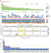
Fig. 1
Fig. 1
Somatic mutations and copy number alterations in pulmonary LELC. a The number of somatic mutations and copy number altered genes for each pulmonary LELC samples in the discovery cohort. Gender, age, smoking status, and tumor stages are listed at the bottom according to the samples. b Signatures are displayed according to the 96-substitution classification, with x-axis showed mutation types and y-axis showed the estimated mutations of each mutation type, which are identified by a Bayesian NMF algorithm. c Frequently mutated genes in the discovery cohort and validation cohort. The two red dashed line denote three and five mutated patients, respectively. Genes mutated in more than five patients are labeled with bold font. LELC, lymphoepithelioma-like carcinoma; SCNA, somatic copy number aberration; NA, not applicable; INDEL, insertion and deletion
Fig. 2
Fig. 2
Analysis of copy number alterations in pulmonary LELC. a Focal amplification and deletion determined from GISTIC 2.0 analysis. The plot shows significant amplification (red) or deletion (blue) for the chromosomes from 1 (top) to 22 (bottom). The green line indicates the cut-off for significance (q = 0.25). Genes listed on left (for amplifications) and right (for deletions) are likely drivers located in the peak areas defined by GISTIC 2.0. b Zoom in the significant deletion region in p21.3 of chromosome 9. Samples are classified into three groups: samples without deletions; samples with heterozygous deletions; and samples with homozygous deletions. c Kaplan–Meier survival analysis for three groups with different copy number status of 9p21.3. Statistical significance was estimated by two-sided log-rank test. LELC, lymphoepithelioma-like carcinoma
Fig. 3
Fig. 3
Altered pathways in pulmonary LELC. Alterations defined as somatic mutations, focal amplifications, and deletions affecting Cell cycle (a), JAK/STAT (b), and NF-kappa B (c) signaling pathways are shown. Alteration frequencies are expressed as a percentage of samples form discovery cohort and validation cohort. LELC, lymphoepithelioma-like carcinoma
Fig. 4
Fig. 4
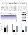
TRAF3 alterations in pulmonary LELC. a Protein domain structure of TRAF3 based on UniProt database with mutated sites. Sequence alignment of TRAF3 protein across distinct species is shown. Amino acid positions of the mutations are indicated above the alignment. b Copy number deletions of TRAF3, samples are sorted by log2 copy number ratio. BEAS-2B cells either with or without knockdown of endogenous TRAF3 expression with short hairpin RNAs (shRNAs) were examined by western blot analysis (c), MTT assay (d), migration assay (e), and colony formation assay (f). Error bars in d, e, and f denote standard error of the mean. Experiments for d, e, and f were performed in triplicate. Student’s t-test was used for statistical analysis of d, e, and f. NC, normal control; MTT, 3-(4, 5-dimethylthiazol-2-yl)-2, 5-diphenyltetrazolium bromide; OD, optical density
Fig. 5
Fig. 5
Comparison between pulmonary LELC and other associated cancer types. a Distribution of non-silent mutation rates of pulmonary LELC and other associated cancer types. The upper numbers indicate the number of samples for each cancer type. Black lines in the boxplot denote median mutation rate for each cancer type and outliers are shown as dots. All the other cancer types are statistically tested by unpaired two-side t-test with pulmonary LELC. *P-value < 0.05 and **P-value < 0.0001. b Mutation spectrum of six mutation type for each cancer type. c Clustering of 96 subtypes based on six mutation types and nucleotides flanking the mutated base for each cancer type. d SCNA comparison of pulmonary LELC and other cancer types. e Frequency comparison of genes with copy number amplification (red) or deletion (blue) for three major oncogenic pathways: NF-kappa B, Cell cycle and Jak/STAT/PI(3)K. Percentages of samples mutated in each cancer type are shown in gray. NPC, nasopharyngeal carcinoma; LELC, lymphoepithelioma-like carcinoma; NKTCL, natural killer/T cell lymphoma; STAD, stomach adenocarcinoma; HNSC, head and neck squamous cell carcinoma; EBV, Epstein-Barr virus; HPV, human papillomavirus; LUAD, lung adenocarcinoma; SCLC, small-cell lung carcinoma; LUSC, lung squamous cell carcinoma; SCNA, somatic copy-number alterations; Mb, megabase

References

    1. Hayashi T, et al. Cytopathologic features and differential diagnostic considerations of primary lymphoepithelioma-like carcinoma of the lung. Diagn. Cytopathol. 2012;40:820–825. doi: 10.1002/dc.21670. - DOI - PubMed
    1. Begin LR, Eskandari J, Joncas J, Panasci L. Epstein-Barr virus related lymphoepithelioma-like carcinoma of lung. J. Surg. Oncol. 1987;36:280–283. doi: 10.1002/jso.2930360413. - DOI - PubMed
    1. Han AJ, Xiong M, Zong YS. Association of Epstein-Barr virus with lymphoepithelioma-like carcinoma of the lung in southern China. Am. J. Clin. Pathol. 2000;114:220–226. doi: 10.1309/148K-ND54-6NJX-NA61. - DOI - PubMed
    1. Travis WD, et al. The 2015 World Health Organization Classification of lung tumors: impact of genetic, clinical and radiologic advances since the 2004 classification. J. Thorac. Oncol. 2015;10:1243–1260. doi: 10.1097/JTO.0000000000000630. - DOI - PubMed
    1. Irie HY, Jaklitsch MT, Shaffer K, Weinstein M, Salgia R. Unusual problems in breast cancer and a rare lung cancer case. Case 4. Primary lymphoepithelioma-like carcinoma of the lung. J. Clin. Oncol. 2003;21:2220–2222. doi: 10.1200/JCO.2003.09.088. - DOI - PubMed

Publication types

MeSH terms