. 2019 Jul 16;9(1):10260.
doi: 10.1038/s41598-019-46727-2.

Cholesterol and 27-hydroxycholesterol promote thyroid carcinoma aggressiveness

Affiliations

Cholesterol and 27-hydroxycholesterol promote thyroid carcinoma aggressiveness

Giovanna Revilla et al. Sci Rep. .

Abstract

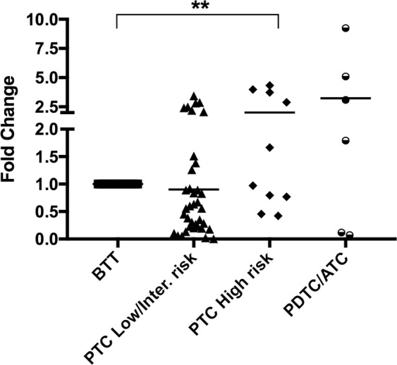
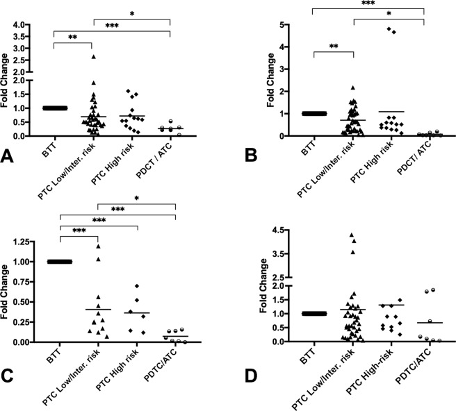
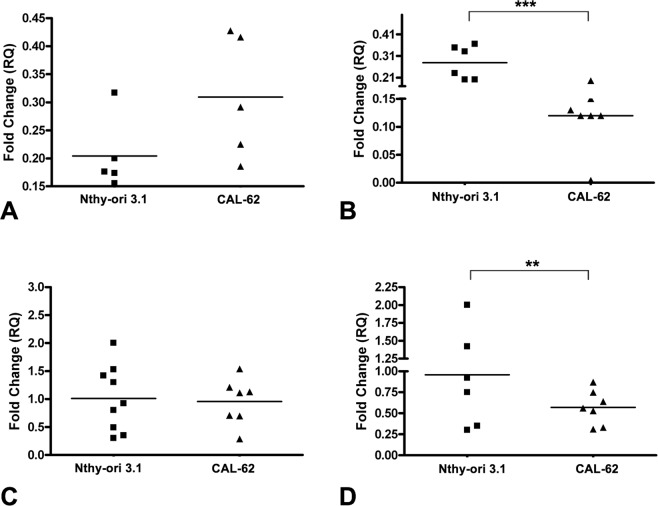
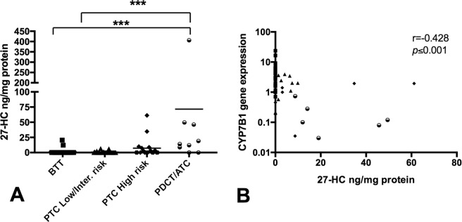
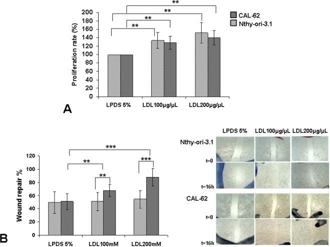
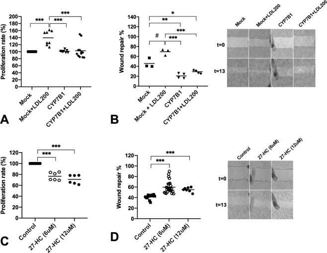
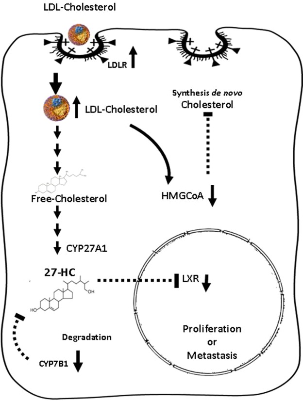
Cholesterol mediates its proliferative and metastatic effects via the metabolite 27-hydroxycholesterol (27-HC), at least in breast and endometrial cancer. We determined the serum lipoprotein profile, intratumoral cholesterol and 27-HC levels in a cohort of patients with well-differentiated papillary thyroid carcinoma (PTC; low/intermediate and high risk), advanced thyroid cancers (poorly differentiated, PDTC and anaplastic thyroid carcinoma, ATC) and benign thyroid tumors, as well as the expression of genes involved in cholesterol metabolism. We investigated the gene expression profile, cellular proliferation, and migration in Nthy-ori 3.1 and CAL-62 cell lines loaded with human low-density lipoprotein (LDL). Patients with more aggressive tumors (high-risk PTC and PDTC/ATC) showed a decrease in blood LDL cholesterol and apolipoprotein B. These changes were associated with an increase in the expression of the thyroid's LDL receptor, whereas 3-hydroxy-3-methylglutaryl-CoA reductase and 25-hydroxycholesterol 7-alpha-hydroxylase were downregulated, with an intratumoral increase of the 27-HC metabolite. Furthermore, LDL promoted proliferation in both the Nthy-ori 3.1 and CAL-62 thyroid cellular models, but only in ATC cells was its cellular migration increased significantly. We conclude that cholesterol and intratumoral accumulation of 27-HC promote the aggressive behavior process of PTC. Targeting cholesterol metabolism could be a new therapeutic strategy in thyroid tumors with poor prognosis.


Conflict of interest statement

The authors declare no competing interests.

Figures

Figure 1
Figure 1
Analysis of the low density lipoprotein receptor (LDLR) gene expression by qRT-PCR in human thyroid tumors. BTT (n = 26), low/intermediate-risk PTC (n = 37), high-risk PTC (n = 10), PDTC/ATC (n = 6). Endogenous expression of the GAPDH gene has been used to normalize the data and BTT as calibrator. Statistical analysis: ANOVA test plus Tukey’s post-test (**p < 0.01).
Figure 2
Figure 2
Analysis of the gene expressions by qRT-PCR in human thyroid tumors. (A) HMGR gene expression, (B) CYP7B1 gene expression, (C) LXR gene expression, (D) CYP27A1 gene expression. The patient cohort was divided into the following groups: BTT (n = 32), low/intermediate-risk PTC (n = 37), high-risk PTC (n = 12), and PDTC/ATC (n = 7). Endogenous expression of the GADPH gene was used to normalize the data, and BTT was used as calibrator tissue. Statistical analysis: ANOVA test plus Tukey’s post-test (*p < 0.05, **p < 0.01, ***p < 0.001).
Figure 3
Figure 3
Analysis of the gene expression by qRT-PCR in thyroid cell lines (Nthy-ori 3.1 and CAL-62). (A) LDLR, (B) HMGR, (C) CYP27A1, and (D) CYP7B1. Endogenous expression of the GADPH gene was used to normalize the data. Statistical analysis: Student t-test (**p < 0.01, ***p < 0.001).
Figure 4
Figure 4
27-hydroxycholesterol (27-HC) content and their relationship with the CYP7B1 gene expression in thyroid tumor tissue extracts. (A) The analysis of 27-HC was measured in 82 patients (n = 97 thyroid tissue samples) corresponding to BTT (n = 40), low/intermediate-risk PTC (n = 30), high-risk PTC (n = 18) and PDTC/ATC (n = 9). The results represent the mean ± SD of the measurements of individual thyroid tissue. (ANOVA) using the Kruskal–Wallis test and Dunn’s post-test (***p < 0.001). (B) A correlation analysis was done in 73 samples in which 27-HC and CYP7B1 expression were analyzed in the same sample (27 BTT, 29 low/intermediate-risk PTC, 11 high-risk PTC and 6 PDTC/ATC samples). The graph illustrates the negative correlation between the CYP7B1 gene expression (2−ΔΔCT) and 27-hydroxycholesterol (27-HC) levels. CYP7B1 gene expression is showed log-transformed. R (−0.428), Pearson’s correlation coefficient; p ≤ 0.001.
Figure 5
Figure 5
Exogenous administration of human low-density lipoprotein (LDL) in thyroid cell lines. (A) Percentage of cellular proliferation of the Nthy-ori 3.1 and CAL-62 cell lines. Both were treated for 24 h with LDL cholesterol (100 μg/mL and 200 μg/mL) compared with control cells maintained in basal conditions (5% LPDS). (B) Monolayer wound-induced migration assay. A line was scratched with a 200-µm plastic pipette tip in CAL-62 and Nthy-ori 3.1 cell lines; cultures were treated for 24 h with LDL cholesterol (100 mg/mL and 200 mg/mL). After 16 h, cells that had migrated to the wounded areas were photographed under a microscope for quantification of cell migration. Images are representative of three separate experiments. Quantitative analysis of wound-induced migration assay compared with control cells maintained in basal conditions (5% LPDS). The results are presented as mean ± SEM of eight experiments done in duplicate. The Kruskal–Wallis test was represented as a ± box plot, n = 8 separate experiments (**p < 0.01, ***p < 0.001).
Figure 6
Figure 6
Overexpression of CYP7B1 gene in CAL-62 cells and exogenous administration of 27-HC in Nthy-ori 3.1 cell. (A) Percentage of cellular proliferation of the CAL-62 cells overexpressing CYP7B1 gene. Cells were treated for 24 h with or without LDL cholesterol (200 μg/mL) compared with control cells maintained in basal conditions (5% FBS). (B) Monolayer wound-induced migration assay in CAL-62 cells overexpressing CYP7B1 gene were scratched with a 200-µm plastic pipette tip and treated for 24 h with or without LDL cholesterol (200 μg/mL). After 13 h the wounded areas were photographed under a microscope for quantification of cell migration. Representative images of experiments. (C) Percentage of cellular proliferation of the Nthy-ori 3.1 cells treated with 27-HC at 6 and 12 μM for 48 h. (D) Monolayer wound-induced migration assay in Nthy-ori 3.1 cells gene were scratched with a 200-µm plastic pipette tip and treated for 48 h with 27-HC at 6 and 12 μM. After 13 h the wounded areas were photographed under a microscope for quantification of cell migration. Statistical analysis: ANOVA test plus Tukey’s post-test (*,#p < 0.05, **p < 0.01, ***p < 0.001).
Figure 7
Figure 7
Schematic drawing of the hypothesis of the effect of uptake and intracellular pathways of LDL in the thyroid cells, based on our results. The LDL cholesterol molecules are internalized via endocytotic vesicles, transported and rapidly metabolized; they start to accumulate in the form of 27-HC due to the inhibition of their degradation in the aggressive forms of tumors. The oxysterol could diffuse into the nucleus region and interact with nuclear receptors and other molecular targets, promoting the proliferation or metastatic processes in the follicular cells. 3-hidroxi-3-metilglutaril-CoA (HMG-CoA) downregulation indicates a potential negative feedback in the de novo synthesis of cholesterol.

References

    1. Martínez-Botas J, et al. Dose-dependent effects of lovastatin on cell cycle progression. Distinct requirement of cholesterol and non-sterol mevalonate derivatives. BBA-Mol Cell Biol. Lipids. 2001;1532:185–194. doi: 10.1016/S1388-1981(01)00125-1. - DOI - PubMed
    1. Fernández C, Lobo M, del VT, Gómez-Coronado D, Lasunción MA. Cholesterol is essential for mitosis progression and its deficiency induces polyploid cell formation. Exp Cell Res. 2004;300:109–120. doi: 10.1016/j.yexcr.2004.06.029. - DOI - PubMed
    1. Rodrigues dos Santos C, et al. LDL- cholesterol signaling induces breast cancer proliferation and invasion. Lipids Health Dis. 2014;13:16. doi: 10.1186/1476-511X-13-16. - DOI - PMC - PubMed
    1. Furuya Y, et al. Low-density lipoprotein receptors play an important role in the inhibition of prostate cancer cell proliferation by statins. Prostate Int. 2016;4:56–60. doi: 10.1016/j.prnil.2016.02.003. - DOI - PMC - PubMed
    1. Sánchez-Martín CC, et al. Cholesterol starvation induces differentiation of human leukemia HL-60 cells. Cancer Res. 2007;67:3379–3386. doi: 10.1158/0008-5472.CAN-06-4093. - DOI - PubMed

Publication types

MeSH terms