. 2019 Jun 1;13(3):271-281.
doi: 10.1302/1863-2548.13.190072.

Challenging clubfeet: the arthrogrypotic clubfoot and the complex clubfoot

Affiliations

Challenging clubfeet: the arthrogrypotic clubfoot and the complex clubfoot

H J P van Bosse. J Child Orthop. .

Abstract

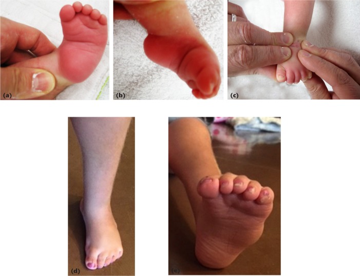
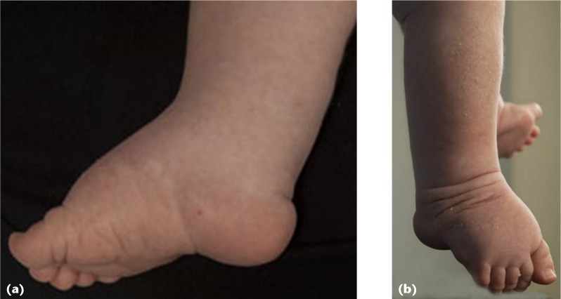
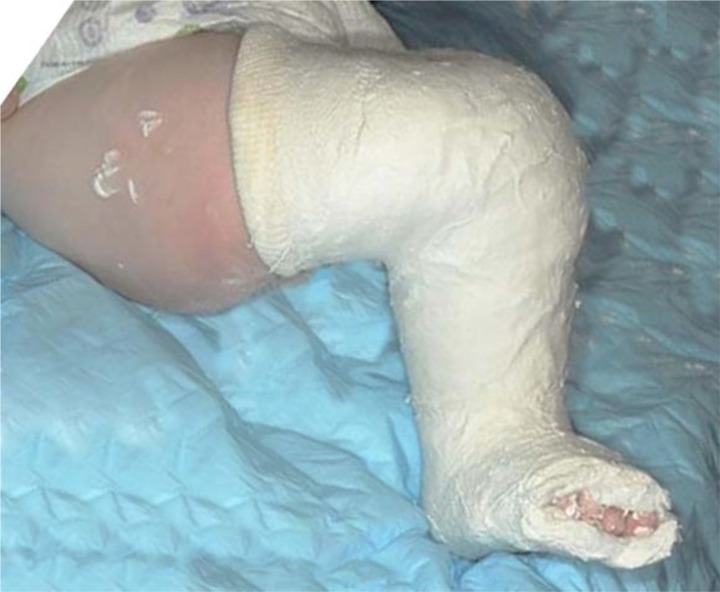
Within the realm of clubfoot deformities, teratologic and complex (or atypical) clubfeet stand out as the most difficult. Exemplarities of the teratologic types of clubfoot are those associated with arthrogryposis multiplex congenita. Treatment of arthrogrypotic clubfoot deformities has been controversial; many different procedures have been advocated, with variable success rates. These clubfeet have a high recurrence rate, regardless of treatment type. Often, the high recurrence rate has led to a high repeat surgery rate, and poor outcomes. Treatment strategies should highlight care that avoids the development of a stiffened foot and allows for a variety of options to regain correction when a relapse occurs. Modifications of the Ponseti method for idiopathic clubfeet have been successful in managing the deformity. The equinocavus variant of the arthrogrypotic clubfoot should be distinguished from the classic clubfoot, as it requires a different treatment method. The equinocavus clubfoot is very similar to the complex or atypical clubfoot. The complex, or atypical, clubfoot also requires a different treatment strategy compared with the typical idiopathic congenital clubfoot. The complex clubfoot appears to be idiopathic in some cases and iatrogenic (due to slipping stretching casts) in others. Dr. Ponseti's modification of his protocol has been effective in treating the deformity. The high recurrence rate suggests the difficulty in maintaining the deformity after correction. The author's preferred treatment for each deformity is included, with an emphasis on minimally invasive methods.

Level of evidence: Level V, expert opinion.

Keywords: Ponseti; arthrogryposis; arthrogryposis multiplex congenita; arthrogrypotic clubfoot; atypical clubfoot; clubfeet; complex clubfoot.


Figures

Fig. 1
Fig. 1
The ‘classic’ arthrogrypotic clubfoot. Both feet of this two-month-old boy have severe heel varus, supination and adductus.
Fig. 2
Fig. 2
Equinocavus clubfoot variant: (a) lateral view of the left foot depicting the severe hindfoot equinus and deep transverse cavus. The toes are mildly flexed in this uncorrected foot; (b) dorsal view of the same foot, showing relatively mild forefoot adductus
Fig. 3
Fig. 3
22-month-old girl with bilateral classic arthrogrypotic clubfeet, had initial and incomplete casting as an newborn: (a) right foot pre-casting anterior view demonstrating the severe adductus; (b) lateral view of the same foot, with severe equinus and cavus; (c) plantar view of the same foot, with severe forefoot adductus and supination and hindfoot varus; (d) plantar view of the right foot, one month later, immediately prior to Achilles tenotomy; (e) lateral view just prior to Achilles tenotomy; (f) lateral view just after tenotomy; after removal of the post-tenotomy cast, 10° of dorsiflexion was possible.
Fig. 4
Fig. 4
Left dual purpose ankle foot orthosis moulded for correction of hindfoot varus and forefoot adductus, the anterior ankle strap originating in the inner lateral aspect of the brace: (a) perspective from directly anterior, demonstrating the straight medial border. Further corrective pads are often placed at the lateral supramalleolar and medial forefoot areas; (b) medial view, with leaf spring ankle visible; (c) anterior perspective with removable dorsiflexion straps in position. The lateral one is tensioned more than the medial, to maintain pronation; (d) medial view with dorsiflexion straps.
Fig. 5
Fig. 5
Equinocavus clubfoot variant in a two-month-old girl: (a) plantar view of left foot, demonstrating medial to lateral plantar crease; (b) medial view showing severe ankle and midfoot equinus, with plantarflexed great toe; (c) the ‘four finger’ technique, the index and long fingers of both hands are positioned over the midfoot to act as a fulcrum, while the thumbs apply dorsiflexion pressure under the heads of the metatarsals. The intact Achilles tendon acts as a counter force, therefore, an Achilles tenotomy should be delayed until full correction of the midfoot cavus; (d) the same foot dorsal view, weight-bearing at seven years’ follow-up; (e) plantar view. The only other treatment she required was percutaneous toe flexor tenotomies at three years of age.
Fig. 6
Fig. 6
three-month-old girl with an idiopathic clubfoot, but with ten previous casts prior to presentation; (a) medial view of right foot, demonstrating high midfoot cavus, deep plantar and posterior ankle ceases and a foreshortened great toe; (b) dorsal view of the same foot with a deep crease at the base of the great toe.
Fig. 7
Fig. 7
Cast moulding to prevent cast slippage. The upper anterior thigh region is made flat, if not actually concave, to prevent kicking of the knee. The proximal tibia is pushed posteriorly on the femur and the anterior border of the tibial section is flat.

References

    1. Ponseti IV, Smoley EN. Congenital club foot: The results of treatment. J Bone Joint Surg [Am] 1963;45-A:261–275.
    1. Ponseti IV. Congenital clubfoot: fundamentals of treatment. New York, NY: Oxford University Press Inc, 1996.
    1. Ponseti IV, Zhivkov M, Davis N, et al. . Treatment of the complex idiopathic clubfoot. Clin Orthop Relat Res 2006;451:171–176. - PubMed
    1. Hall JG. Arthrogryposis multiplex congenita: etiology, genetics, classification, diagnostic approach, and general aspects. J Pediatr Orthop B 1997;6:159–166. - PubMed
    1. Bevan WP, Hall JG, Bamshad M, et al. . Arthrogryposis multiplex congenita (amyoplasia): an orthopaedic perspective. J Pediatr Orthop 2007;27:594–600. - PubMed