. 2019 Jun 1;13(3):293-303.
doi: 10.1302/1863-2548.13.190079.

Treatment of relapsed, residual and neglected clubfoot: adjunctive surgery

Affiliations

Treatment of relapsed, residual and neglected clubfoot: adjunctive surgery

M Eidelman et al. J Child Orthop. .

Abstract

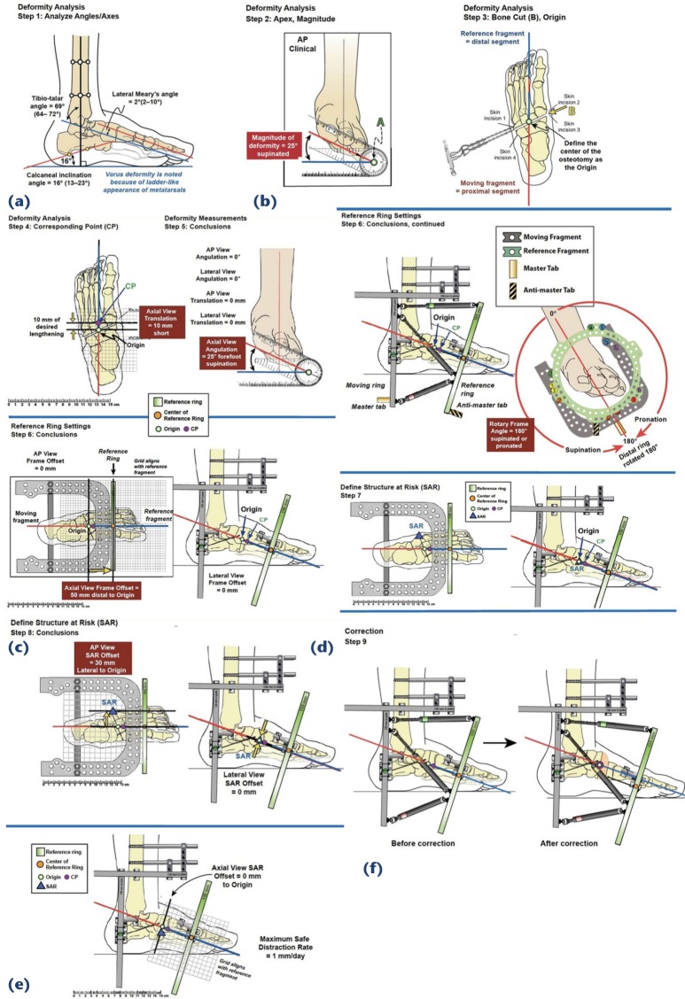
Over the past two decades, the Ponseti 'conservative' (non-surgical) method of clubfoot treatment has been almost universally adopted worldwide. As a result, the need for operative treatment for clubfoot has decreased dramatically. However, even Ponseti himself routinely used surgery for certain patients: at least 90% of feet need percutaneous tenotomy, and 15% to 40% may require tibialis anterior tendon transfer. Additionally, relapses are common, sometimes necessitating further surgical intervention. Relapses are recurrent deformities in previously well corrected feet. Residual deformities may be defined as persistent deformities in incompletely corrected feet. In addition, in many parts of the developing world, neglected clubfoot is still a major challenge. Many neglected feet can be treated with Ponseti principles, particularly in younger children. However, in older children and adults, surgical approaches are more likely to be needed. Major reasons for relapsed/residual clubfoot include incomplete application of the Ponseti principles, inability to adhere to the foot abduction brace protocol, failure to recommend a complete course of bracing and inadequate follow-up. Sometimes, despite excellent treatment, and perfect adherence to the bracing protocols, there are still relapses, related to intrinsic muscle imbalance. We describe several solutions that include reinstitution of Ponseti casting and 'á la carte' operative treatment. As an alternative for particularly stubborn cases, application of a hexapod external fixator can be a powerful tool. In order to be a full-service clubfoot specialist, and not only a Ponseti practitioner, one must have in their toolbox the full gamut of adjunctive surgical options.

Level of evidence: V.

Keywords: clubfoot; neglected; relapsed; residual.


Figures

Fig. 1
Fig. 1
(a) Four-year-old boy with relapsed clubfoot, lost to follow-up and noncompliant with foot abduction brace (printed with permission); (b) foot after four Ponseti casts and before tibialis anterior transfer.
Fig. 2
Fig. 2
The ‘Ponse-Taylor’ hexapod strategy to correct residual/relapsed clubfoot (used with permission, Rubin Institute for Advanced Orthopedics, Sinai Hospital of Baltimore). Worm’s eye view: before and after derotation to correct the internal spin through the subtalar joint. Anteroposterior (AP) view: during the initial varus to valgus correction and derotation, the talar neck wire is attached to the tibial ring with step down plates, to focus the correction through the subtalar joint. Next the talar neck wire is transferred to the foot ring, and then the equinus correction is focused on the ankle joint. Lateral view: the gradual correction of the equinus to neutral, and then additional over correction into approximately 20° dorsiflexion, anticipating some rebound.
Fig. 3
Fig. 3
Ponse-Taylor II: used for cases in which there is also a forefoot adductus or cavus (used with permission, Rubin Institute for Advanced Orthopedics, Sinai Hospital of Baltimore). This is the last step, after the internal rotation, varus and hindfoot equinus have been corrected. The foot ring is cut on both sides, removing a segment from the lateral side in order to allow the cut ends to shorten. Threaded rods are applied over one-hole posts to act as a distractor on the medial side, and to allow compression (neutralization) on the lateral side, to correct adductus. For pure cavus, both sides may be distracted.
Fig. 4
Fig. 4
Hexapod ‘Butt’ frame to correct residual forefoot supinatus/adductus. This assumes that the hindfoot is well aligned (neutral) (used with permission, Rubin Institute for Advanced Orthopedics, Sinai Hospital of Baltimore): (a) lateral view of foot showing normal hindfoot orientation, but supinatus of the forefoot; (b) anteroposterior (AP) view of foot demonstrates forefoot supinatus. Percutaneous Gigli saw cut through the cuneiforms and cuboid; (c) the forefoot is designated as the reference segment, and the (green) ring applied perpendicular to that segment but rotated to reflect the supinatus (25°). The designated origin and corresponding point which are about 10 mm apart to permit disengagement of the osteotomy, to make it easier to rotate, can correct the deformity; (d) the forefoot is the reference segment, so it is depicted parallel to the floor, and the rest of foot (the proximal portion) is offset in an angular fashion. The structure at risk (SAR) is shown as the lateral edge of the osteotomy which is the farthest point from the centre of rotation (middle of the foot); (e) the offsets of the SAR are shown measured on the different views; (f) lateral view of the foot before and after correction. The foot is initially supinated, and then flat.
Fig. 5
Fig. 5
A 16-year-old boy with neglected clubfoot. Printed courtesy of Dr Scott Nelson, Port au Prince, Haiti.
Fig. 6
Fig. 6
Miter Taylor Spatial Frame correction of neglected clubfoot.

References

    1. Dobbs MB, Morcuende JA, Gurnett CA, Ponseti IV. Treatment of idiopathic clubfoot: an historical review. Iowa Orthop J 2000;20:59-64. - PMC - PubMed
    1. Wenger DR, Rang M The art and practice of children orthopedics. New York: Raven Press, 1992.
    1. Ponseti IV, Smoley EN. Congenital club foot: the results of treatment. J Bone Joint Surg [Am] 1963;45-A:261-344.
    1. Ponseti IV. Congenital clubfoot: fundamentals of treatment. New York: Oxford University Inc, 1996.
    1. Coplan JA, Herzenberg JE. Non-operative treatment of congenital clubfoot : McCarthy JJ, Drennan JC The child’s foot and ankle (2nd edition). Philadelphia: Lippincott Williams &Wilkins, 2010:64-74.