

The application of convolutional neural network to stem cell biology
- PMID: 31312276
- PMCID: PMC6611022
- DOI: 10.1186/s41232-019-0103-3
The application of convolutional neural network to stem cell biology
Abstract
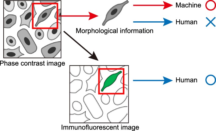
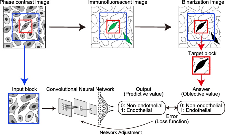
Induced pluripotent stem cells (iPSC) are one the most prominent innovations of medical research in the last few decades. iPSCs can be easily generated from human somatic cells and have several potential uses in regenerative medicine, disease modeling, drug screening, and precision medicine. However, further innovation is still required to realize their full potential. Machine learning is an algorithm that learns from large datasets for pattern formation and classification. Deep learning, a form of machine learning, uses a multilayered neural network that mimics human neural circuit structure. Deep neural networks can automatically extract features from an image, although classical machine learning methods still require feature extraction by a human expert. Deep learning technology has developed recently; in particular, the accuracy of an image classification task by using a convolutional neural network (CNN) has exceeded that of humans since 2015. CNN is now used to address several tasks including medical issues. We believe that CNN would also have a great impact on the research of stem cell biology. iPSCs are utilized after their differentiation to specific cells, which are characterized by molecular techniques such as immunostaining or lineage tracing. Each cell shows a characteristic morphology; thus, a morphology-based identification system of cell type by CNN would be an alternative technique. The development of CNN enables the automation of identifying cell types from phase contrast microscope images without molecular labeling, which will be applied to several researches and medical science. Image classification is a strong field among deep learning tasks, and several medical tasks will be solved by deep learning-based programs in the future.
Keywords: Artificial intelligence; Deep learning; Endothelial cell; Image recognition; Induced pluripotent stem cell; Machine learning; Stem cell.
Conflict of interest statement
Competing interestsSY owns equity in Heartseed, Inc.
Figures

References
-
- Takahashi K, Tanabe K, Ohnuki M, Narita M, Ichisaka T, Tomoda K, et al. Induction of pluripotent stem cells from adult human fibroblasts by defined factors. Cell. 2007;131(5):861–872. - PubMed
-
- Takahashi K, Yamanaka S. Induction of pluripotent stem cells from mouse embryonic and adult fibroblast cultures by defined factors. Cell. 2006;126(4):663–676. - PubMed
-
- Nagoshi N, Okano H. Applications of induced pluripotent stem cell technologies in spinal cord injury. J Neurochem. 2017;141(6):848–860. - PubMed
-
- Yuasa S, Fukuda K. Recent advances in cardiovascular regenerative medicine: the induced pluripotent stem cell era. Expert Rev Cardiovasc Ther. 2008;6(6):803–810. - PubMed
-
- Yuasa S, Fukuda K. Cardiac regenerative medicine. Circulation. 2008;72(Supplement A):A49–A55. - PubMed
Publication types
LinkOut - more resources
Full Text Sources
