. 2019 Jun 15;11(6):3384-3397.
eCollection 2019.

MiR-9 functions as a tumor suppressor in acute myeloid leukemia by targeting CX chemokine receptor 4

Affiliations

MiR-9 functions as a tumor suppressor in acute myeloid leukemia by targeting CX chemokine receptor 4

Bingke Zhu et al. Am J Transl Res. .

Abstract

MicroRNAs (miRNAs) play key roles in the pathogenesis of many cancers, including acute myeloid leukemia (AML). Although miRNA-9 (miR-9) is involved in the leukemogenesis of AML, the underlying mechanisms remain to be elucidated. In this study, we found that miR-9 and C-X-C chemokine receptor 4 (CXCR4) were differentially expressed in myeloid leukemia, particularly in AML. The inverse correlation between miR-9 and CXCR4 was identified in AML samples and cell lines. The AML patients simultaneously with high levels of CXCR4 and low expression of miR-9 possessed poor prognosis. In vitro, miR-9 inhibited the proliferation, apoptosis resistance, migration, and invasion of AML cells. Dual luciferase assays verified CXCR4 as a direct target of miR-9. The suppressive effects of miR-9 on AML cells were counteracted or mimicked by CXCR4 overexpression or depletion, respectively. Overall, this study reveals that miR-9 retards the aggressive behaviors of AML cells by repressing CXCR4. Thus, miR-9/CXCR4 axis may represent a potential therapeutic target for AML.

Keywords: C-X-C chemokine receptor 4; acute myeloid leukemia; apoptosis; growth; microRNA-9.


Conflict of interest statement

None.

Figures
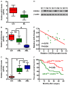
Figure 1
Figure 1
Expression of miR-9 and CXCR4 in AML samples. (A) qPCR assay was performed to determine the miR-9 levels in BM samples of ML patients and healthy individuals (control). U6 was used as the normal control. (B and C) Comparisons of the miR-9 expression (B) and CXCR4 mRNA level (C) in BM samples of the control, CML, and AML patients. U6 and GAPDH were used as the endogenous controls, respectively. (D) The protein level of CXCR4 in BM samples of the control and AML patients was measured by Western blot assay. β-actin were used as the normal control. (E) The inverse relation with miR-9 levels and CXCR4 protein levels in AML samples. (F) Five-year overall survival rate of AML patients with miR-9 low expression and CXCR4 high level (n = 25) and the patients with miR-9 high expression and CXCR4 low level (n = 11). Data are presented as mean ± SD of three independent experiments. *P < 0.05. AML: acute myeloid leukemia; CML: chronic myeloid leukemia; ML: myeloid leukemia; N: normal samples; T: AML samples; NS: Not significant.
Figure 2
Figure 2
Levels of miR-9 and CXCR4 in AML cells. (A) qPCR assay was conducted to detect the miR-9 levels in AML cell lines (NB4, HL-60, Kasumi-1, SKNO-1, and KG-1a) and the normal CD34+ cells. U6 was used as the normal control. (B and C) The mRNA expression (B) and protein level (C) of CXCR4 in AML cells and CD34+ cells. GAPDH and β-actin were used as the normal controls, respectively. (D) The relationship between miR-9 expression and protein levels of CXCR4 in AML cells were evaluated by Pearson’s correlation analysis. Data are presented as mean ± SD of three independent experiments. *P < 0.05 compared with CD34+ cells.
Figure 3
Figure 3
MiR-9 inhibited the proliferation, migration, and invasion and promoted apoptosis of AML cells. Kasumi-1 and SKNO-1 cells with or without 10 µg/mL of VCR pretreatment were transfected with 100 nM of miR-NC or miR-9 mimics. (A) CCK-8 assay was carried out to assess cell proliferation at 24, 48, 72, and 96 h after transfection. (B and C) Cell cycle distribution (B) and cell apoptosis (C) were analyzed by flow cytometry at 48 h after transfection. (D and E) Transwell assays were performed to evaluate the migration (D) and invasion (E) of cells at 48 h after transfection. Data are presented as mean ± SD of three independent experiments. *P < 0.05, **P < 0.01 compared with miR-NC group. VCR: Vincristine.
Figure 4
Figure 4
MiR-9 directly targeted CXCR4 in AML cells. (A) Three classical softwares were used to predict that miR-9 directly targets CXCR4. (B) Predicted binding site in 3’-UTR of CXCR4 for miR-9. (C) Dual luciferase assays were carried out after Kasumi-1 and SKNO-1 cells were co-transfected pGL3-luc-WT 3’-UTR-CXCR4 or pGL3-luc-MUT 3’-UTR-CXCR4 with miR-9 or miR-NC mimics for 48 h. mRNA (D) and protein (E) levels of CXCR4 in Kasumi-1 and SKNO-1 cells were measured by qPCR and Western blot assays. GAPDH and β-actin were used as internal controls, respectively. Data are presented as mean ± SD of three independent experiments. *P < 0.05 compared with miR-NC group.
Figure 5
Figure 5
CXCR4 overexpression attenuated the anti-leukemic activity of miR-9 in AML cells. Kasumi-1 and SKNO-1 cells with or without 10 µg/mL of VCR pretreatment were transfected with miR-NC or miR-9 or miR-9 + CXCR4-expressing plasmid. (A) Cell proliferation was measured by CCK-8 assay at 24, 48, 72, and 96 h after transfection. (B and C) Cell cycle (B) and cell apoptosis (C) was analyzed by flow cytometrys at 48 h after transfection. (D and E) Transwell assays were performed to evaluate the migration (D) and invasion (E) of cells at 48 h after transfection. (F) Western blot results of CXCR4, Ki67, cyclin D1, Bax, Bcl-2, MMP-2, and MMP-9 expression at 48 h after transfection. β-actin was used as the normal control. Data are presented as mean ± SD of three independent experiments. *P < 0.05, **P < 0.01 compared with miR-NC + VCR (-) group, #P < 0.05 compared with miR-9 + VCR (-) group, §P < 0.05 compared with miR-9 + VCR (-) group, ‖P < 0.05 compared with miR-NC + VCR (+) group, ◆P < 0.05 compared with miR-9 + CXCR4 + VCR (-) group, ●P < 0.05 compared with miR-9 + VCR (+) group. VCR: Vincristine.
Figure 6
Figure 6
Overexpression of miR-9 or knockdown of CXCR4 impaired AML growth in vivo. SCID mice were subcutaneously injected with Kasumi-1-luc cells infected with control lentivirus (Lenti-psi-CHECK-2 or Lenti-pLKO.1) or a recombinant lentivirus expressing miR-9 precursor (Lenti-psi-CHECK-2-miR-9) or shCXCR4 (Lenti-shCXCR4). A. In vivo luciferase image for detection of xenograft tumor growth in mice implanted with various cells. B. Tumor volume was monitored once a week for 5 weeks. C. Tumor weight was measured after 5 weeks of implantation. D. TUNEL assay was conducted to determine the percentage of apoptotic cells. E. Western blot results of CXCR4, Ki67, cyclin D1, Bax, Bcl-2, MMP-2, and MMP-9 expression in different groups. β-actin was used as the normal control. Data are presented as mean ± SD of three independent experiments. *P < 0.05 vs. Lenti-psi-CHECK-2 or Lenti-pLKO.1 group; **P < 0.01 vs. Lenti-psi-CHECK-2 or Lenti-pLKO.1 group.

References

    1. Lowenberg B, Downing JR, Burnett A. Acute myeloid leukemia. N Engl J Med. 1999;341:1051–1062. - PubMed
    1. Estey E, Dohner H. Acute myeloid leukaemia. Lancet. 2006;368:1894–1907. - PubMed
    1. Estey EH. Treatment of acute myeloid leukemia. Haematologica. 2009;94:10–16. - PMC - PubMed
    1. Buyse M, Michiels S, Squifflet P, Lucchesi KJ, Hellstrand K, Brune ML, Castaigne S, Rowe JM. Leukemia-free survival as a surrogate end point for overall survival in the evaluation of maintenance therapy for patients with acute myeloid leukemia in complete remission. Haematologica. 2011;96:1106–1112. - PMC - PubMed
    1. Ambros V. The functions of animal microRNAs. Nature. 2004;431:350–355. - PubMed

LinkOut - more resources