

Inadvertent excision of malignant soft tissue tumours
- PMID: 31312520
- PMCID: PMC6598609
- DOI: 10.1302/2058-5241.4.180060
Inadvertent excision of malignant soft tissue tumours
Abstract
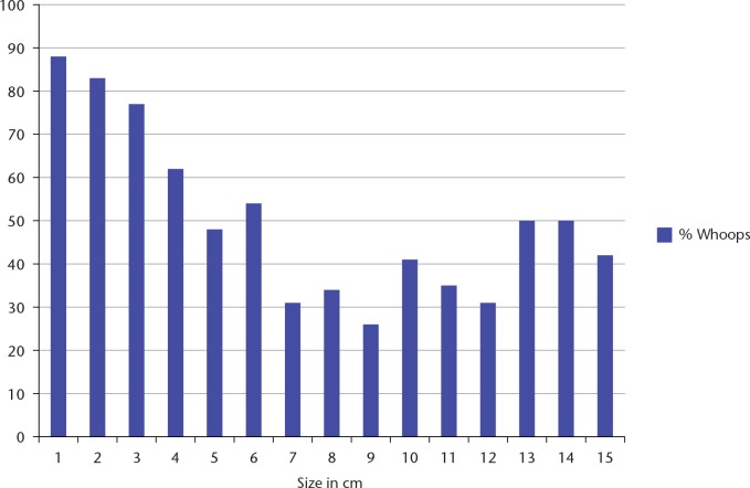
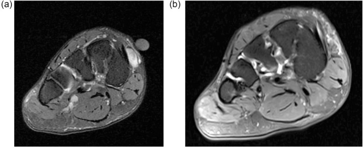
Inadvertent excision of lumps which turn out to be sarcomas is not uncommon.Imaging has a limited role in detecting microscopic residual disease but can show the extent of the previous surgical field.Standard treatment is wide re-excision, usually combined with radiotherapy.Residual tumour is found in an average of 50% of reported cases.The presence of residual disease is an adverse prognostic factor.All lumps bigger than a golf ball should have a diagnosis prior to excision. Cite this article: EFORT Open Rev 2019;4 DOI: 10.1302/2058-5241.4.180060.
Keywords: inadvertent excision; local recurrence; soft tissue sarcoma; unplanned excision; wide re-excision.
Conflict of interest statement
ICMJE Conflict of interest statement: None declared.
Figures
References
-
- Giuliano AE, Eilber FR. The rationale for planned reoperation after unplanned total excision of soft-tissue sarcomas. J Clin Oncol 1985;3:1344–1348. - PubMed
-
- Gustafson P, Dreinhöfer KE, Rydholm A. Soft tissue sarcoma should be treated at a tumor center: a comparison of quality of surgery in 375 patients. Acta Orthop Scand 1994;65:47–50. - PubMed
-
- von Mehren M, Randall RL, Benjamin RS, et al. Soft tissue sarcoma, version 2.2018, NCCN clinical practice guidelines in oncology. J Natl Compr Canc Netw 2018;16:536–563. - PubMed
Publication types
LinkOut - more resources
Full Text Sources
