. 2019 Jun 24;4(3):404-421.
doi: 10.1016/j.jacbts.2019.02.004. eCollection 2019 Jun.

Western Diet-Fed, Aortic-Banded Ossabaw Swine: A Preclinical Model of Cardio-Metabolic Heart Failure

Affiliations

Western Diet-Fed, Aortic-Banded Ossabaw Swine: A Preclinical Model of Cardio-Metabolic Heart Failure

T Dylan Olver et al. JACC Basic Transl Sci. .

Abstract

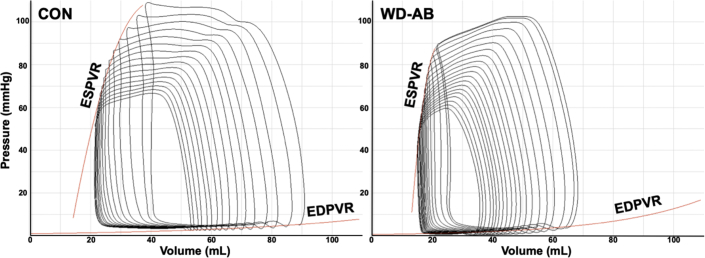
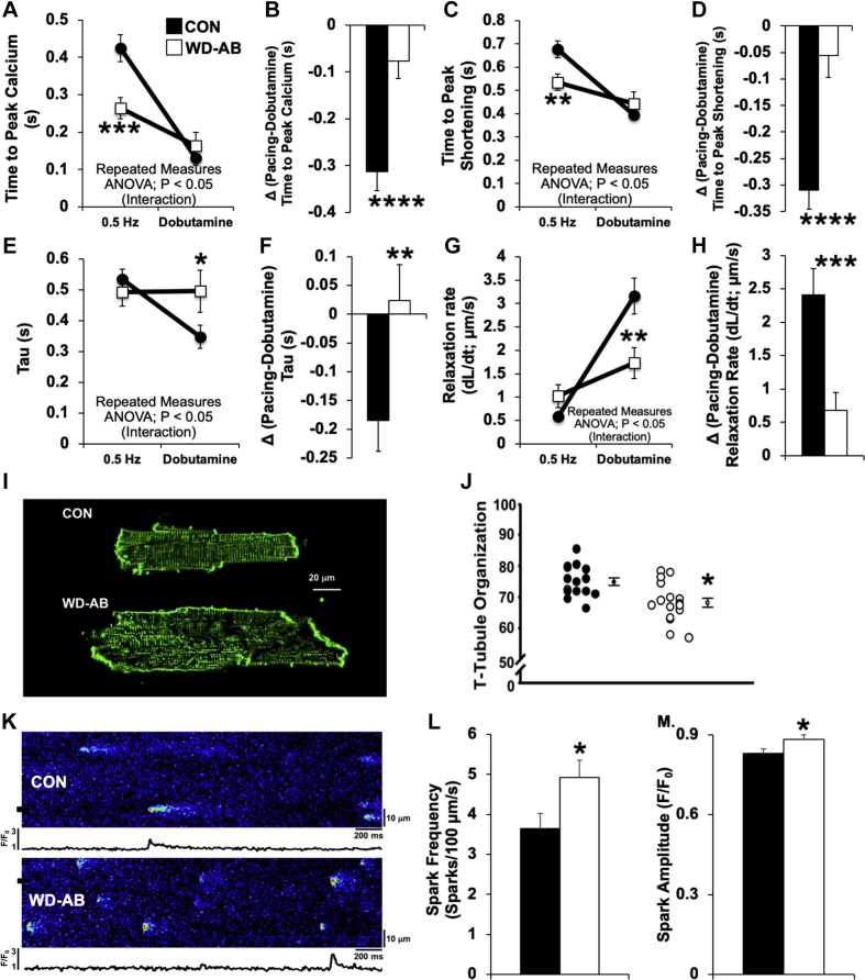
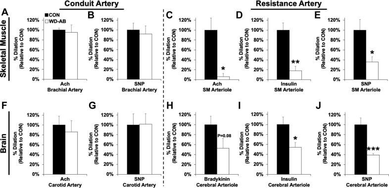
The development of new treatments for heart failure lack animal models that encompass the increasingly heterogeneous disease profile of this patient population. This report provides evidence supporting the hypothesis that Western Diet-fed, aortic-banded Ossabaw swine display an integrated physiological, morphological, and genetic phenotype evocative of cardio-metabolic heart failure. This new preclinical animal model displays a distinctive constellation of findings that are conceivably useful to extending the understanding of how pre-existing cardio-metabolic syndrome can contribute to developing HF.

Keywords: AB, aortic-banded; CON, control; EDPVR, end-diastolic pressure−volume relationship; EF, ejection fraction; HF, heart failure; HFpEF, heart failure with preserved ejection fraction; HFrEF, heart failure with reduced ejection fraction; IL1RL1, interleukin 1 receptor-like 1; LV, left ventricle; NF, nuclear factor; PTX3, pentraxin-3; WD, Western Diet; cardio-metabolic disease; heart failure; integrative pathophysiology; preclinical model of cardiovascular disease.


Figures

None
Graphical abstract
Figure 1
Figure 1
Representative P-V Loops Representative pressure−volume (P-V) loops at rest from individual control (CON) and Western diet, aortic-banded (WD-AB) animals. EDPVR = end-diastolic P-V relationship; ESPVR = end-systolic P-V relationship.
Figure 2
Figure 2
Ossabaw Swine Fed A WD With Chronic Pressure Overload-Induced HF Show Impaired Individual Cardiomyocyte Function (A) Systolic cardiomyocyte calcium and (C) shortening kinetics are faster under baseline pacing conditions in the WD-AB group, but (B and D) lack β-adrenergic reserve in response to dobutamine. (E and F) Diastolic calcium reuptake (tau) and (G and H) relaxation rate kinetic reserve following exposure to dobutamine is impaired in WD-AB cardiomyocytes. (I) Representative cardiomyocyte images show (J) cardiomyocyte t-tubule disorganization in WD-AB animals. (K) Representative line scans illustrate spontaneous ryanodine receptor-mediated calcium (L) spark frequency and (M) amplitude were increased in the WD-AB group. *t-test versus CON (*p < 0.05; **p < 0.01; ***p < 0.001; ****p < 0.0001). n = 4 animals, 25 to 27 cells in the CON group; n = 4 animals, 23 to 25 cells in the WD-AB group. ANOVA = analysis of variance; HF = heart failure; other abbreviations as in Figure 1.
Figure 3
Figure 3
Isolated Mitochondrial Function Is Compromised in Ossabaw Swine Fed a WD With Chronic Pressure Overload-Induced HF and Associated With Decreased L4CL (A) Mitochondrial dysfunction evident as impaired complex 1 and 2-dependent respiration and functional uncoupling of the respiratory chain and adenosine triphosphate synthesis. (B to E) Susceptibility to calcium (Ca2+)-induced mitochondrial permeability transition (an early indicator of mitochondrial dysfunction) was increased in the WD-AB group. (B and C) Quantification of the area above the curve of the Ca2+-induced swelling traces was increased in WD-AB animals. (D and E) Conversely, Ca2+-retention capacity was decreased in the WD-AB group. (F to H) The composition of (G) tetralinoleoyl cardiolipin (L4CL) to (F) total cardiolipin levels was (H) decreased in WD-AB animals. *t-test versus CON (*p < 0.05; ** p < 0.01). n = 4 for CON and WD-AB groups. ADP = adenosine diphosphate; MG = malate/glutamate; other abbreviations as in Figures 1 and 2.
Figure 4
Figure 4
Ossabaw Swine Fed a WD With Chronic Pressure Overload-Induced HF Exhibit Distinct Molecular Signatures Indicative of LV Pathological Remodeling (A) Left ventricular (LV) gene interactions between significant cardiac hypertrophy and cardiac fibrosis networks revealed by ingenuity pathway analysis between CON and WD-AB animals. (B) Pentraxin-3 (PTX3) mRNA level is increased in the WD-AB group and (C) negatively correlated with LV untwisting. WD-AB animals show a right and downward shift along this relationship compared with CON (+ = individual animal data points forming the regression line). (D) Interleukin 1 receptor-like 1 (IL1RL1) mRNA level was also increased in the WD-AB group and (E) negatively correlated with LV untwisting. WD-AB animals again show a right and downward shift along this relationship compared with CON (+ = individual animal data points forming the regression line). (F) The collagen I/III mRNA ratio is increased in the WD-AB group. (G) A decrease in the more compliant N2BA titin isoform is seen in WD-AB animals with representative samples from both the CON and WD-AB groups presented alongside the bar graph. *t-test versus CON (*p < 0.05). n = 3 for CON and WD-AB in Figure 4A. n = 5 for the CON group and n = 4 for the WD-AB group in Figures 4B to 4G. Abbreviations as in Figures 1 and 2.
Figure 4
Figure 4
Ossabaw Swine Fed a WD With Chronic Pressure Overload-Induced HF Exhibit Distinct Molecular Signatures Indicative of LV Pathological Remodeling (A) Left ventricular (LV) gene interactions between significant cardiac hypertrophy and cardiac fibrosis networks revealed by ingenuity pathway analysis between CON and WD-AB animals. (B) Pentraxin-3 (PTX3) mRNA level is increased in the WD-AB group and (C) negatively correlated with LV untwisting. WD-AB animals show a right and downward shift along this relationship compared with CON (+ = individual animal data points forming the regression line). (D) Interleukin 1 receptor-like 1 (IL1RL1) mRNA level was also increased in the WD-AB group and (E) negatively correlated with LV untwisting. WD-AB animals again show a right and downward shift along this relationship compared with CON (+ = individual animal data points forming the regression line). (F) The collagen I/III mRNA ratio is increased in the WD-AB group. (G) A decrease in the more compliant N2BA titin isoform is seen in WD-AB animals with representative samples from both the CON and WD-AB groups presented alongside the bar graph. *t-test versus CON (*p < 0.05). n = 3 for CON and WD-AB in Figure 4A. n = 5 for the CON group and n = 4 for the WD-AB group in Figures 4B to 4G. Abbreviations as in Figures 1 and 2.
Figure 5
Figure 5
Ossabaw Swine Fed a WD With Chronic Pressure Overload-Induced HF Demonstrate Skeletal Muscle and Brain Vascular Dysfunction in Isolated Microvessels Brachial artery ring preparations indicate (A) endothelial and (B) smooth muscle−dependent function is similar between the CON and WD-AB groups. Skeletal muscle microvessel preparations show significant (C) endothelial, (D) metabolic, and (E) smooth muscle−dependent dysfunction in the WD-AB group. Carotid artery ring preparations reveal (F) endothelial and (G) smooth muscle−dependent function is similar between the CON and WD-AB group. Cerebral microvessel preparations show significant (H) endothelial, (I) metabolic, and (J) smooth muscle−dependent dysfunction in WD-AB animals. *t-test versus CON (*p < 0.05; **p < 0.01; ***p < 0.001). n = 5 for the CON group, and n = 4 for the WD-AB group. Ach = acetylcholine; SM = skeletal muscle; SNP = sodium nitroprusside; other abbreviations as in Figures 1 and 2.
Figure 6
Figure 6
Ossabaw Swine Fed a WD With Chronic Pressure Overload-Induced HF Exhibit a Systemic Inflammatory State Consistent With Common Comorbidities Seen in Experimental HF Animals in the WD-AB group were (A) obese, (B) inactive, and (C) insulin resistant (homeostatic model assessment of insulin resistance [HOMA-IR]). Dyslipidemia was observed in WD-AB animals as indicated by (D) increased plasma cholesterol, (E) triglycerides, (F) low-density lipoproteins (LDL), and (G) 2-chlorostearate. (H and I) Plasma liver enzymes were increased and evidence of (J; Masson’s trichrome stain) increased liver fibrosis were seen in the WD-AB group. *t-test versus CON (*p < 0.05; **p < 0.01; ***p < 0.001). n = 5 for CON and WD. Abbreviations as in Figures 1 and 2.

References

    1. Borlaug B.A., Paulus W.J. Heart failure with preserved ejection fraction: pathophysiology, diagnosis, and treatment. Eur Heart J. 2011;32:670–679. - PMC - PubMed
    1. Maeder M.T., Kaye D.M. Heart failure with normal left ventricular ejection fraction. J Am Coll Cardiol. 2009;53:905–918. - PubMed
    1. Oktay A.A., Rich J.D., Shah S.J. The emerging epidemic of heart failure with preserved ejection fraction. Curr Heart Fail Rep. 2013;10:401–410. - PMC - PubMed
    1. Zhao Z., Wang H., Jessup J.A., Lindsey S.H., Chappell M.C., Groban L. Role of estrogen in diastolic dysfunction. Am J Physiol Heart Circ Physiol. 2014;306:H628–H640. - PMC - PubMed
    1. Sharma K., Kass D.A. Heart failure with preserved ejection fraction: mechanisms, clinical features, and therapies. Circ Res. 2014;115:79–96. - PMC - PubMed