. 2019 Nov;256(6):1705-1726.
doi: 10.1007/s00709-019-01412-z. Epub 2019 Jul 17.

Comparative studies on structure of the floral nectaries and the abundance of nectar production of Prunus laurocerasus L

Affiliations

Comparative studies on structure of the floral nectaries and the abundance of nectar production of Prunus laurocerasus L

Mirosława Chwil et al. Protoplasma. 2019 Nov.

Abstract

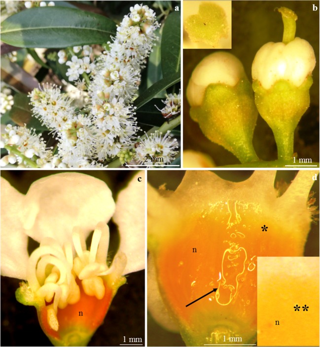
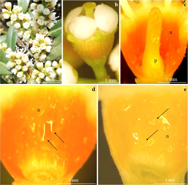
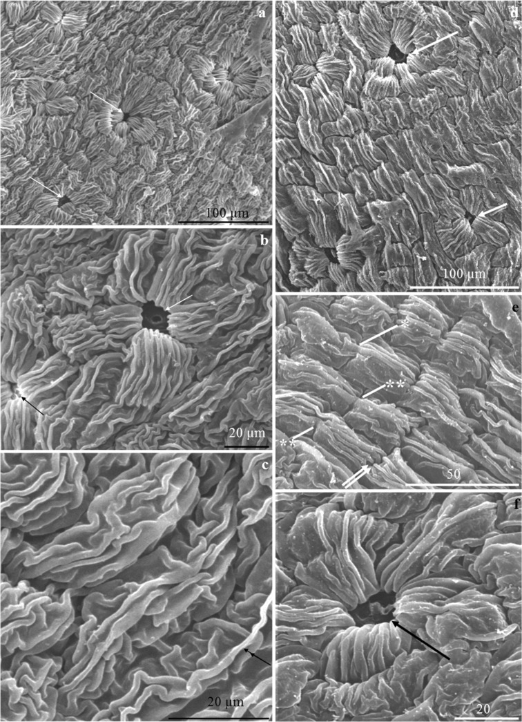
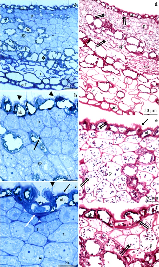
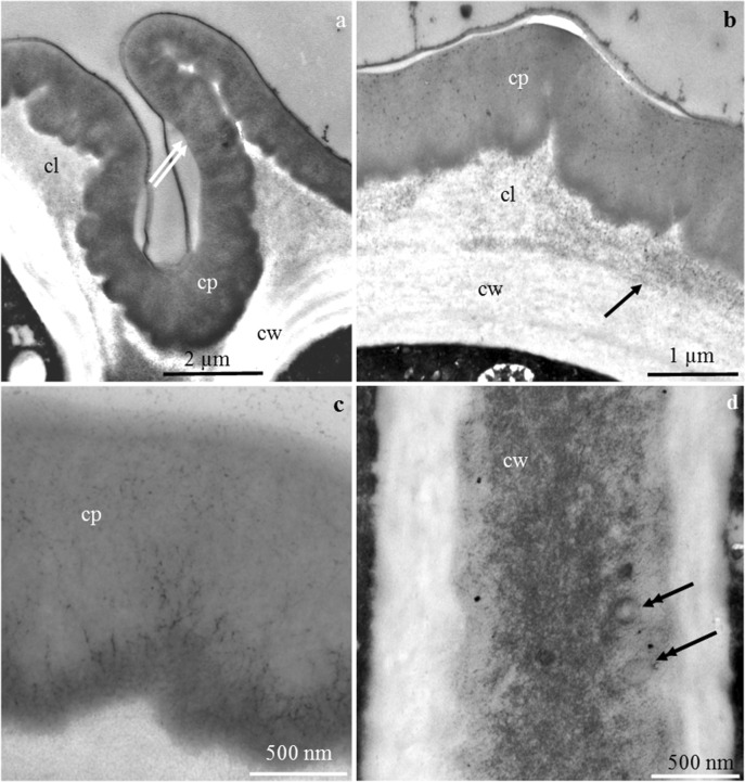
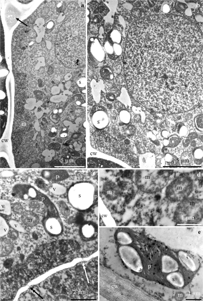
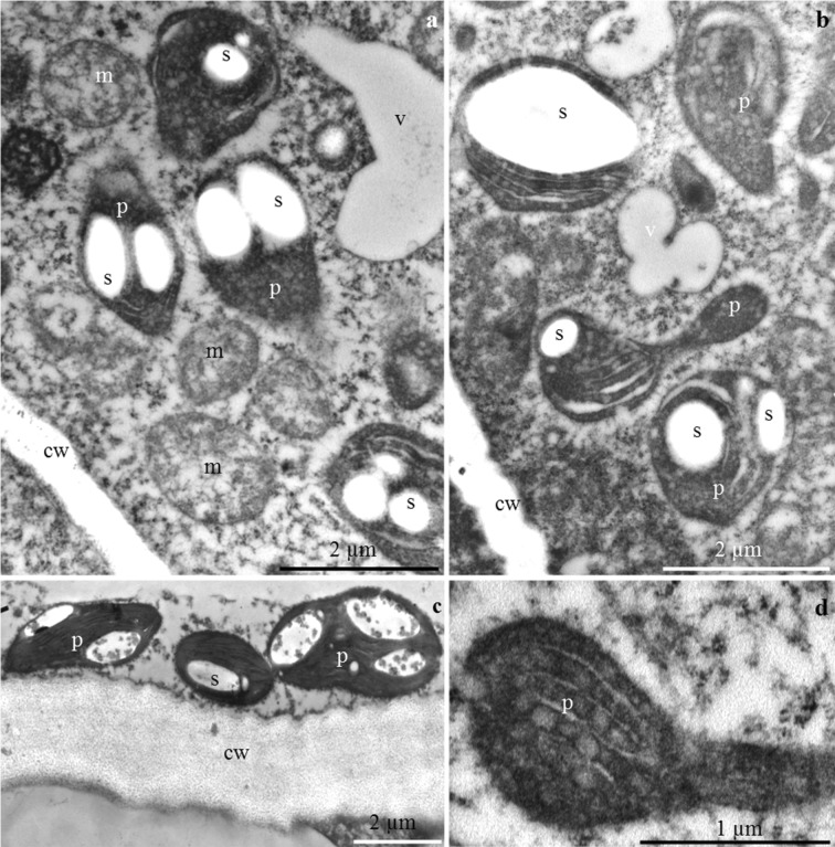
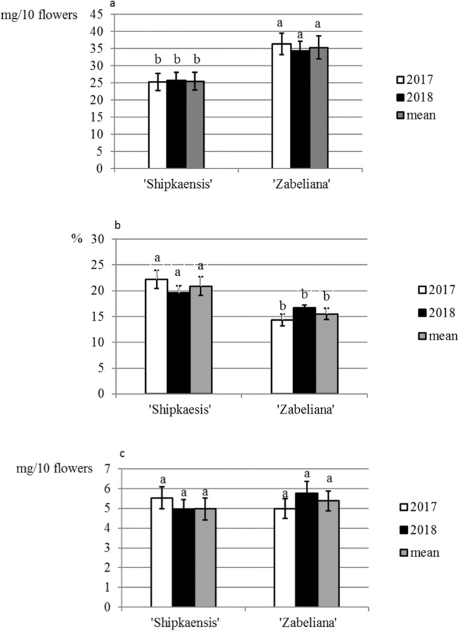
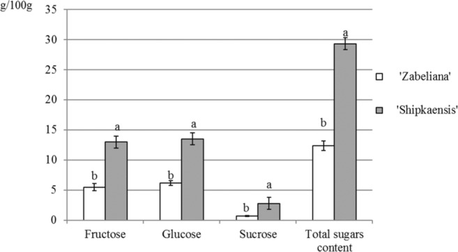
There is very scanty information concerning the floral nectary structure and nectar secretion in Prunus laurocerasus L. Therefore, the aim of the study was to determine the micromorphology, anatomy and ultrastructure of nectaries; the abundance of nectar production; and the quantitative and qualitative composition of sugars contained in the nectar of two P. laurocerasus cultivars: 'Schipkaensis' and 'Zabeliana'. The nectary structure was studied using light, fluorescence, scanning and transmission electron microscopy techniques. The nectar sugars were analysed with HPLC. The 'Schipkaensis' had longer inflorescences with a larger number of flowers and a longer perianth than 'Zabeliana'. The micromorphological structure of the nectaries in 'Schipkaensis' exhibited denser (approx. 39%) and larger (approx. 50%) stomata and thicker (approx. 13%) cuticular striae forming wider bands (approx. 26%) than in 'Zabeliana'. The results provide new data on the micromorphology, anatomy and ultrastructure of these floral nectaries. Nectary cuticle ornamentation as well as the size, type and density of stomata and stomatal complex topography can have a diagnostic value in Prunus. The nectar sugar weight indicates a significant apicultural value of the cherry laurel, especially in the case of 'Schipkaensis'. Cherry laurel is an entomophilous species recommended for cultivation in nectariferous zones and insect pollinator refuges; however, climatic conditions eliminating the invasiveness of these plants should be considered.

Keywords: Anatomy; Cherry laurel; Floral biology; Micromorphology; Sugar components; Ultrastructure.


Conflict of interest statement

The authors declare that they have no conflict of interest.

Figures

Fig. 1
Fig. 1
a–dPrunus laurocerasus ‘Schipkaensis’ inflorescence, flowers, and nectary. a Raceme with buds in different developmental phases. b Bursting buds with visible protogyny. c Bursting bud (onset of nectar secretion), visible intense orange nectary (n). d Bright orange-yellow (asterisk) and yellow colour (two asterisks) of the nectary (n) at a later stage of nectar secretion, visible nectar (arrow). b SM. c, d LM
Fig. 2
Fig. 2
a–ePrunus laurocerasus ‘Zabeliana’ inflorescence (a), flowers (b) and nectary (ce). a Raceme with buds in different developmental phases. b Bursting bud with visible protogyny. c Intense orange colour of the nectary (n) at the beginning of nectar secretion (bursting bud), visible upper pistil (p). d Small and large drops of nectar (arrow) on the nectary surface (n) in the flowers at the onset of anthesis, visible intense orange colour of the active secretory cells. e Bright yellow colour of the nectary (n) in a blooming flower (full nectar secretion phase), visible accumulated nectar (arrow). b SM. ce LM
Fig. 3
Fig. 3
a–fPrunus laurocerasus ‘Schipkaensis’ (ac) and ‘Zabeliana’ (df) nectary epidermis. a Striated cuticular ornamentation, visible stomata (arrow) evenly distributed on the nectary surface. b Open stomata (arrow), located below other nectary epidermis cells, visible radial arrangement of cells in the stomatal complex with parallel or intertwined arrangement of cuticular striae. c Corrugated cuticular striae arranged along the longer cell axis of other epidermis cells sometimes with long major striae (double-headed arrow). d Striated cuticular ornamentation, visible stomata (arrow). e Cuticular striae in parallel arrangement sometimes with a slight bend maintaining continuity (asterisk) with the wall or forming a bend (two asterisks) over the anticlinal wall or convergent with the anticlinal wall (two arrows), visible striation along the longer axis of the cells. f Radial pattern of cuticular striae on the surface of guard cells. SEM
Fig. 4
Fig. 4
a–c Surface of the epidermis (a) and cross sections of the nectary (b, c) in P. laurocerasus ‘Schipkaensis’ (a, c) and ‘Zabeliana’ (b). a Stomatal complex and other epidermis cells, visible autofluorescence of cuticular striae (arrow). b Intense autofluorescence (double-headed arrow) of the nectary epidermis (ab), visible nectary parenchyma (p) and sub-nectary parenchyma (sp) cells, vascular bundle (vb) located on the border between the sub-nectary parenchyma and receptacle (r). c Epicuticular fluorescence (two arrows) of the nectary epidermis (ab). FM
Fig. 5
Fig. 5
a–f Longitudinal sections of receptacle (a, d) and nectary cells (af) in P. laurocerasus ‘Schipkaensis’(a, d, f) and ‘Zabeliana’ (b, c, e). a Epidermal nectary cells (ab), tightly arranged nectary parenchyma cells (p), a layer of larger cells of sub-nectary parenchyma (sp), loosely arranged receptacle cells (pr). b Epidermal nectary cells (ab) with convexities of the outer cell wall (arrowhead), with more intensely coloured cytoplasm and various degrees of vacuolisation, nectary parenchyma cells (p) with dense cytoplasm, large nucleus and one or two nucleoli, large sub-nectary parenchyma cells (sp) with one or two small vacuoles, visible precipitates in the cell sap (triple-headed arrow). c Thick outer cell wall of epidermis (arrow), stomata (double-headed arrow) located below other epidermis cells, numerous vesicular structures in the nectary parenchyma cells (p), large centrally located nucleus (n) with nucleoli. d Pink stained cell wall polysaccharides, amyloplasts (two arrows) in the epidermal (ab) and parenchyma (p) nectary cells, visible vascular bundles (vb). e Intensely stained nectary epidermis cell walls, convexities (arrow) in the outer epidermis walls, amyloplasts (two arrows) in the parietal epidermis cytoplasm and numerous amyloplasts in the nectary parenchyma cells (p). f Amyloplasts (two arrows) in guard cells (s), in other epidermis cells (ab) and nectary parenchyma (p), visible protuberances of the outer cell wall (arrow). LM
Fig. 6
Fig. 6
a–d The outer cell wall of nectary epidermis in P. laurocerasus ‘Schipkaensis’ (a, c) and ‘Zabeliana’ (b, d). a Outer periclinal cell wall, visible convexities of the wall (two arrows), lamellar (cp) and reticulate (cl) cuticle and the remaining part of the wall (cw). b Cuticle band uniformly distributed on the other part of the wall, visible lamellar (cp) and reticulate (cl) cuticle and pectin band (arrow). c Lamellar band of cuticle (cp). d Inner periclinal cell wall, visible vesicles in the wall (double-headed arrow). TEM
Fig. 7
Fig. 7
af. Nectary epidermis cells in P. laurocerasus ‘Schipkaensis’. a, b Electron dense protoplast, visible numerous plastids (p) with starch grains (s), mitochondria (m), spherical nucleus (n), and vesicular structures (arrow) in extracellular spaces (a). c Dense cytoplasm, visible pleomorphic plastids (p) with starch grains (s), mitochondria (m) located near the wall and plastids, small vacuoles (v), plasmodesmata in the periclinal cell wall (arrow with double arrowhead). d Mitochondria clustered near the cell wall (cw). e Plastid (p) with starch grains (s) located at the cell wall (cw), visible mitochondrion (m). TEM
Fig. 8
Fig. 8
ad Nectary parenchyma cells in P. laurocerasus ‘Schipkaensis’. a, b Numerous plastids (p) with starch grains (s), mitochondria (m) located close to amyloplasts and cell walls (cw), small vacuole (v). c Mitochondria (m), pleomorphic plastids (p) with starch grains (s), cell wall (cw). d Elongated plastid (p). TEM
Fig. 9
Fig. 9
ad Nectary epidermis cells in P. laurocerasus ‘Zabeliana’. a Thick outer cell wall (cw), visible protoplast with numerous starch grains (s) and small vacuoles (v). b Dense cytoplasm, numerous mitochondria (m) and plastids (p) with starch grains (s). c Plasmodesmata (two arrows) in the anticlinal cell wall (cw), large cell nucleus (n) with dense nucleoplasm, mitochondria (m) located close to the cell wall and nucleus, plastids (p) with starch grains (s). d Anticlinal cell wall (cw), visible vesicles (v) in the intercellular spaces (arrow with double arrowhead), clustered mitochondria (m). TEM
Fig. 10
Fig. 10
ad Nectary parenchyma cells in P. laurocerasus ‘Zabeliana’. a Electron dense cytoplasm, plastids (p) with starch grains (s), numerous mitochondria (m), spherical cell nucleus (n), radially arranged ER. b Elongated cell nucleus (n), visible nucleolus, pleomorphic mitochondria (m) located at the cell wall and nucleus. c Serially arranged mitochondria (m) close to the anticlinal cell wall (cw). d Cell nucleus (n), coupled mitochondria (m), visible rough reticulum (RER). TEM
Fig. 11
Fig. 11
ac Mass of nectar (a), concentration of sugars in nectar (b) and mass of sugar in nectar (c) in the studied cultivars of P. laurocerasus. Explanations: Mean values for each feature of nectar secretion abundance, calculated from twelve replications (n = 12), marked with the same letter are not different at P < 0.05 based on the HSD Tukey test. Vertical bars represent the standard deviation (SD) of means
Fig. 12
Fig. 12
Total sugar content and percentage of sugars: glucose, fructose and sucrose in the nectar of the two cultivars P. laurocerasus studied. Explanations: Mean values for quantitative and qualitative composition of sugars in the nectar, calculated from three replications (n = 3), marked with the same letter are not different at P ≤ 0.05 based on the HSD Tukey test. Vertical bars represent the standard deviation (SD) of means

Similar articles

Cited by

References

    1. Bernardello GA. A systematic survey of floral nectaries. In: Nicolson M, Nepi E, Pacini E, editors. Nectaries and nectar. Dordrecht: Springer; 2007. pp. 19–127.
    1. Bogdanov S, Martin P, Lüllmann C, Borneck R, Flamini Ch, Morlot MCh, Heretier J, Vorwohl G, Russmann H, Persano-Oddo L, Sabatini AG, Marcazzan GL, Marioleas P, Tsigouri K, Kerkvliet J, Ortiz A, Ivanov T (1997) Harmonised methods of the European honey commission. Apidologie Spec Iss 28:1–59
    1. Bordács MM, Botz L, Orosz-Kovács Z, Kerek MM. The composition of nectar in apricot cultivars. Acta Hortic. 1995;384:367–371. doi: 10.17660/ActaHortic.1995.384.58. - DOI
    1. Bukovics P, Orosz-Kovács Z, Szabó LG, Farkas Á, Bubán T. Composition of floral nectar and its seasonal variability in sour cherry cultivars. Acta Bot Hungar. 2003;45:259–271. doi: 10.1556/ABot.45.2003.3-4.2. - DOI
    1. Cameron RW, Taylor JE, Emmett NR. What’s ‘cool’ in the world of green façades? How plant choice influences the cooling properties of green walls. Build Environ. 2014;73:198–207. doi: 10.1016/j.buildenv.2013.12.005. - DOI

MeSH terms

LinkOut - more resources