. 2019 Sep 1;122(3):1213-1225.
doi: 10.1152/jn.00264.2019. Epub 2019 Jul 17.

Estradiol rapidly modulates excitatory synapse properties in a sex- and region-specific manner in rat nucleus accumbens core and caudate-putamen

Affiliations

Estradiol rapidly modulates excitatory synapse properties in a sex- and region-specific manner in rat nucleus accumbens core and caudate-putamen

Amanda A Krentzel et al. J Neurophysiol. .

Abstract

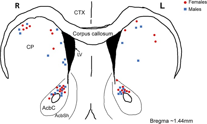
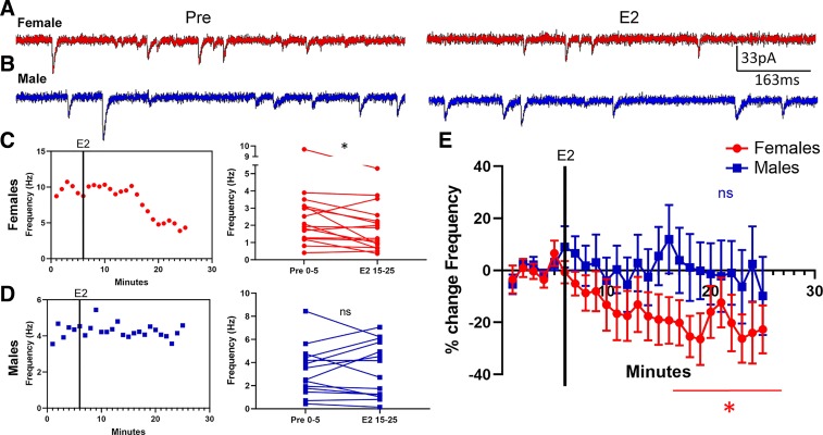
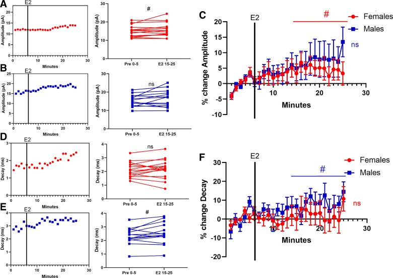
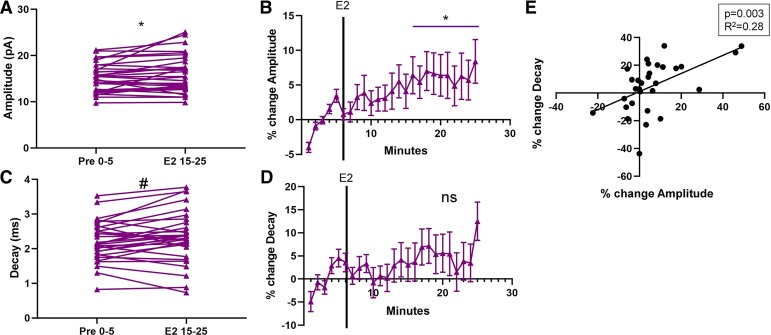
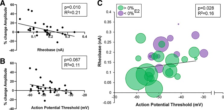
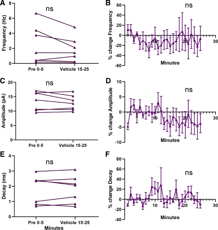
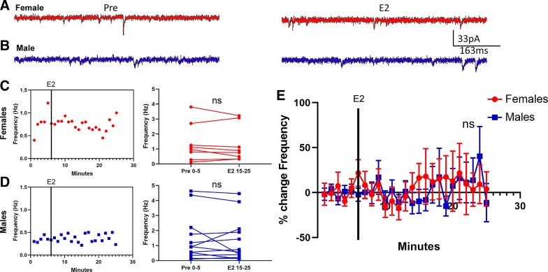
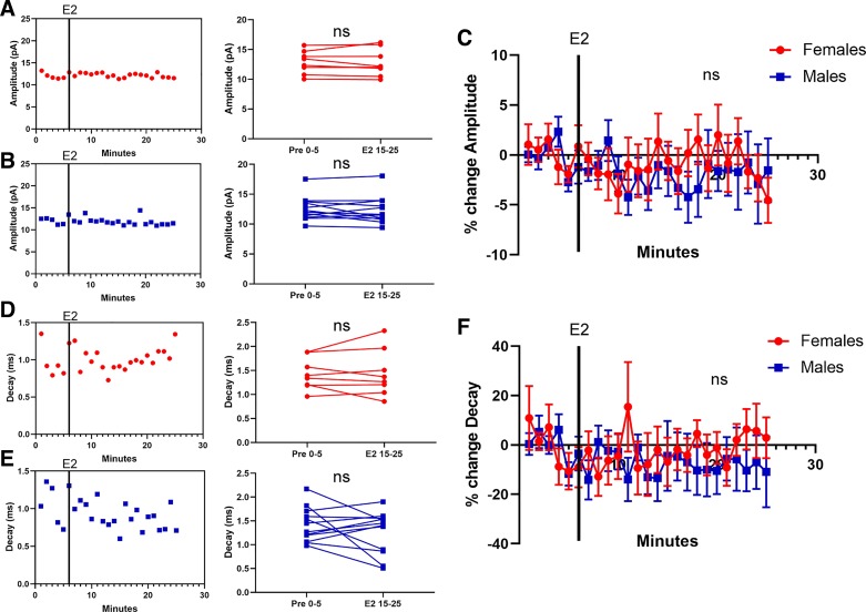
Estradiol acutely facilitates sex differences in striatum-dependent behaviors. However, little is understood regarding the underlying mechanism. In striatal regions in adult rodents, estrogen receptors feature exclusively extranuclear expression, suggesting that estradiol rapidly modulates striatal neurons. We tested the hypothesis that estradiol rapidly modulates excitatory synapse properties onto medium spiny neurons (MSNs) of two striatal regions, the nucleus accumbens core and caudate-putamen in adult female and male rats. We predicted there would be sex-specific differences in pre- and postsynaptic locus and sensitivity. We further analyzed whether MSN intrinsic properties are predictive of estrogen sensitivity. Estradiol exhibited sex-specific acute effects in the nucleus accumbens core: miniature excitatory postsynaptic current (mEPSC) frequency robustly decreased in response to estradiol in female MSNs, and mEPSC amplitude moderately increased in response to estradiol in both male and female MSNs. This increase in mEPSC amplitude is associated with MSNs featuring increased intrinsic excitability. No MSN intrinsic electrical property associated with changes in mEPSC frequency. Estradiol did not acutely modulate mEPSC properties in the caudate-putamen of either sex. This is the first demonstration of acute estradiol action on MSN excitatory synapse function. This demonstration of sex and striatal region-specific acute estradiol neuromodulation revises our understanding of sex hormone action on striatal physiology and resulting behaviors.NEW & NOTEWORTHY This study is the first to demonstrate rapid estradiol neuromodulation of glutamatergic signaling on medium spiny neurons (MSNs), the major output neuron of the striatum. These findings emphasize that sex is a significant biological variable both in MSN sensitivity to estradiol and in pre- and postsynaptic mechanisms of glutamatergic signaling. MSNs in different regions exhibit diverse responses to estradiol. Sex- and region-specific estradiol-induced changes to excitatory signaling on MSNs explain sex differences partially underlying striatum-mediated behaviors and diseases.

Keywords: caudate-putamen; estradiol; medium spiny neuron; miniature excitatory postsynaptic currents; nucleus accumbens; striatum.


Conflict of interest statement

No conflicts of interest, financial or otherwise, are declared by the authors.

Figures

Fig. 1.
Fig. 1.
Location of whole cell patch-clamped medium spiny neurons (MSNs) in adult female and male rat nucleus accumbens core (AcbC) and caudate-putamen (CP). Blue squares are MSNs recorded from males; red circles are MSNs recorded from females. AbcSh, nucleus accumbens shell; CTX, cortex; L, left hemisphere; LV, lateral ventricle; R, right hemisphere.
Fig. 2.
Fig. 2.
Estradiol rapidly decreased miniature excitatory postsynaptic current (mEPSC) frequency in female rat nucleus accumbens core (AcbC) medium spiny neurons (MSNs). A and B: representative examples of mEPSCs recorded in female (A) and male (B) MSNs before (Pre; left) and during exposure to 17β-estradiol (E2; right). C and D: an example experiment in females (C) and males (D) demonstrating the time course of the estradiol-induced decrease in mEPSC frequency (left) and a plot illustrating mean mEPSC frequency during a 5-min baseline before and during estradiol exposure (right). Connected symbols indicate data from an individual neuron. E: plotting the normalized change in mEPSC frequency vs. time indicates that female AcbC MSNs (n = 16) are more sensitive to rapid estradiol action than are male AcbC MSNs (n = 14). Red circles indicate females and blue squares indicate males. Black vertical lines in CE depict the time point at which estradiol entered the bath. E2 15–25; the last 10 min during 17β-estradiol exposure; Pre 0–5, the 5-min baseline period before estradiol exposure. *P < 0.05; ns, not statistically significant.
Fig. 3.
Fig. 3.
Estradiol, miniature excitatory postsynaptic current (mEPSC) amplitude, and mEPSC decay in female or male rat nucleus accumbens core (AcbC) medium spiny neurons (MSNs). A and B: an example experiment in females (A) and males (B) demonstrating the time course of the estradiol-induced increase in mEPSC amplitude (left) and a plot illustrating mean mEPSC amplitude before and during estradiol exposure (right). Connected symbols indicate data from an individual neuron. C: plotting the normalized change in mEPSC amplitude disaggregated by sex. D and E: an example experiment in females (D) and males (E) demonstrating the time course of the estradiol-induced increase in mEPSC decay (left) and a plot illustrating mean mEPSC decay before and during estradiol exposure (right). Connected symbols indicate data from an individual neuron. F: normalized changes or lack thereof in mEPSC decay in females (n = 16) or males (n = 14). Red circles indicate females and blue squares indicate males. Black vertical lines depict the time point in which estradiol entered the bath. E2 15–25; the last 10 min during 17β-estradiol exposure; Pre 0–5, the 5-min baseline period before estradiol exposure. #P < 0.10; ns, not statistically significant.
Fig. 4.
Fig. 4.
Estradiol rapidly increases miniature excitatory postsynaptic current (mEPSC) amplitude in rat nucleus accumbens core (AcbC) medium spiny neurons (MSNs) independently of sex. A: mean mEPSC amplitude before and during estradiol exposure in both males and females (n = 30). Connected symbols indicate data from an individual neuron. B: plotting the normalized change in mEPSC amplitude in both males and females indicates that both sexes are sensitive to the effects of estradiol. C: mean mEPSC decay before and during estradiol exposure in both males and females. D: plotting the normalized change in mEPSC decay in both males and females indicates little effect of estradiol. E: relationship of %change in amplitude to %change in decay in both male and female AcbC MSNs. Purple triangles indicate both sexes together. Black line is the linear relationship, and there is a significant positive correlation between %change in amplitude and %change in decay. E2 15–25; the last 10 min during 17β-estradiol exposure; Pre 0–5, the 5-min baseline period before estradiol exposure.*P < 0.05; #P < 0.10; ns, not statistically significant.
Fig. 5.
Fig. 5.
Sensitivity of miniature excitatory postsynaptic current (mEPSC) amplitude to estradiol correlates with neuronal excitability in both sexes in rat nucleus accumbens core (AcbC) medium spiny neurons (MSNs). A: relationship of rheobase to the %change in mEPSC amplitude in response to estradiol. B: relationship of action potential threshold to the %change in mEPSC amplitude in response to estradiol. C: combined relationship of rheobase and action potential threshold to the %change in mEPSC amplitude in response to estradiol. Green circles indicate increased %changes of mEPSC amplitude in response to estradiol. Purple circles indicate decreased %changes of mEPSC amplitude in response to estradiol. Increasing circle size indicates larger %change. Black lines indicate the best-fit linear relationship.
Fig. 6.
Fig. 6.
Miniature excitatory postsynaptic current (mEPSC) parameters do not change with vehicle exposure in rat nucleus accumbens core (AcbC) medium spiny neurons (MSNs). A: plot illustrating no effect of vehicle exposure on mEPSC frequency in males and females (n = 7). Connected symbols indicate data from an individual neuron. B: normalized mEPSC frequency. C: plot illustrating no effect of vehicle exposure on mEPSC amplitude in males and females. D: normalized mEPSC amplitude. E: plot illustrating no effect of vehicle exposure on mEPSC decay in males and females. F: normalized mEPSC decay. Purple triangles indicate both sexes together. E2 15–25; the last 10 min during 17β-estradiol exposure; ns, not statistically significant; Pre 0–5, the 5-min baseline period before estradiol exposure.
Fig. 7.
Fig. 7.
Estradiol does not modulate miniature excitatory postsynaptic current (mEPSC) frequency in female or male caudate-putamen (CP) medium spiny neurons (MSNs). A: representative examples of mEPSCs recorded in female MSNs before (Pre; left) and during exposure to 17β-estradiol (E2; right). B: representative examples of mEPSCs recorded in male MSNs before (left) and during exposure to estradiol (right). C and D: an example experiment in females (C) and males (D) demonstrating the time course of no changes in mEPSC frequency in response to estradiol (left) and a plot illustrating mean mEPSC frequency during a 5-min baseline before and during estradiol exposure (right). Connected symbols indicate data from an individual neuron. E: plotting the normalized change in mEPSC frequency vs. time indicates that neither female (n = 8) nor male (n = 12) MSN mEPSC frequency is sensitive to rapid estradiol exposure. Red circles indicate females and blue squares indicate males. Black vertical lines depict the time point at which estradiol entered the bath. E2 15–25; the last 10 min during 17β-estradiol exposure; Pre 0–5, the 5-min baseline period before estradiol exposure; ns, not statistically significant.
Fig. 8.
Fig. 8.
Estradiol does not modulate miniature excitatory postsynaptic current (mEPSC) amplitude or decay in female or male caudate-putamen (CP) medium spiny neurons (MSNs). A and B: an example experiment in females (A) and males (B) demonstrating the time course of no changes in mEPSC amplitude in response to estradiol (left) and a plot illustrating mean mEPSC amplitude before and during estradiol exposure (right). Connected symbols indicate data from an individual neuron. C: plotting the normalized change in mEPSC amplitude disaggregated by sex reveals no sensitivity to estradiol exposure. D and E: an example experiment in females (n = 8; D) and males (n = 12; E) demonstrating the time course of no changes in mEPSC decay in response to estradiol (left) and a plot illustrating mean mEPSC decay before and during estradiol exposure (right). F: plotting the normalized change in mEPSC decay disaggregated by sex reveals no sensitivity to estradiol exposure. Red circles indicate females and blue squares indicate males. Black vertical lines depict the time point at which estradiol entered the bath. E2 15–25; the last 10 min during 17β-estradiol exposure; Pre 0–5, the 5-min baseline period before estradiol exposure; ns, not statistically significant.

Similar articles

Cited by

References

    1. Adams C, Chen X, Moenter SM. Changes in GABAergic transmission to and intrinsic excitability of gonadotropin-releasing hormone (GnRH) neurons during the estrous cycle in mice. eNeuro 5: ENEURO.0171-18.2018, 2018. doi:10.1523/ENEURO.0171-18.2018. - DOI - PMC - PubMed
    1. Adams C, DeFazio RA, Christian CA, Milescu LS, Schnell S, Moenter SM. Changes in both neuron intrinsic properties and neurotransmission are needed to drive the increase in GnRH neuron firing rate during estradiol-positive feedback. J Neurosci 39: 2091–2101, 2019. doi:10.1523/JNEUROSCI.2880-18.2019. - DOI - PMC - PubMed
    1. Almey A, Filardo EJ, Milner TA, Brake WG. Estrogen receptors are found in glia and at extranuclear neuronal sites in the dorsal striatum of female rats: evidence for cholinergic but not dopaminergic colocalization. Endocrinology 153: 5373–5383, 2012. doi:10.1210/en.2012-1458. - DOI - PMC - PubMed
    1. Almey A, Milner TA, Brake WG. Estrogen receptors in the central nervous system and their implication for dopamine-dependent cognition in females. Horm Behav 74: 125–138, 2015. doi:10.1016/j.yhbeh.2015.06.010. - DOI - PMC - PubMed
    1. Almey A, Milner TA, Brake WG. Estrogen receptor α and G-protein coupled estrogen receptor 1 are localized to GABAergic neurons in the dorsal striatum. Neurosci Lett 622: 118–123, 2016. doi:10.1016/j.neulet.2016.04.023. - DOI - PMC - PubMed

Publication types